中国神经再生研究(英文版) ›› 2025, Vol. 20 ›› Issue (10): 3013-3024.doi: 10.4103/NRR.NRR-D-23-01651

• 原著:神经损伤修复保护与再生 • 上一篇    下一篇

新冠肺炎睡眠障碍患者存在持续性灰质结构异常:一项为期3个月的纵向研究


  • 出版日期:2025-10-15 发布日期:2025-02-09

Persistent alterations in gray matter in COVID-19 patients experiencing sleep disturbances: a 3-month longitudinal study

Kaixuan Zhou1, 2, #, Gaoxiong Duan2, #, Ying Liu2 , Bei Peng2 , Xiaoyan Zhou2 , Lixia Qin3 , Lingyan Liang2 , Yichen Wei2 , Qingping Zhang2 , Xiaocheng Li2 , Haixia Qin2 , Yinqi Lai2 , Yian Lu3 , Yan Zhang2 , Jiazhu Huang2 , Jinli Huang2 , Yinfei Ouyang2 , Bolin Bin2 , Mingming Zhao3 , Jun Liu4, *, Jianrong Yang5, *, Demao Deng1, 2, *   

  1. 1 Guangxi Key Laboratory of Special Biomedicine; School of Medicine, Guangxi University, Nanning, Guangxi Zhuang Autonomous Region, China;  2 Department of Radiology, the People’s Hospital of Guangxi Zhuang Autonomous Region, Guangxi Academy of Medical Sciences, Nanning, Guangxi Zhuang Autonomous Region, China;  3 Department of Sleep Medicine, the People’s Hospital of Guangxi Zhuang Autonomous Region, Nanning, Guangxi Zhuang Autonomous Region, China;  4 Department of Radiology, The Second Xiangya Hospital, Central South University, Changsha, Hunan Province, China;  5 Guangxi Clinical Research Center for Sleep Medicine, the People’s Hospital of Guangxi Zhuang Autonomous Region, Nanning, Guangxi Zhuang Autonomous Region, China
  • Online:2025-10-15 Published:2025-02-09
  • Contact: Demao Deng, MD, demaodeng@163.com; Jianrong Yang, MBBS, gandansurgery2014@163.com; Jun Liu, MD, junliu123@csu.edu.cn.
  • Supported by:
    This study was supported by grants from Major Project of Science and Technology of Guangxi Zhuang Autonomous Region, No. Guike-AA22096018 (to JY); Guangxi Key Research and Development Program, No. AB22080053 (to DD); Major Project of Science and Technology of Guangxi Zhuang Autonomous Region, No. Guike-AA23023004 (to MZ); the National Natural Science Foundation of China, Nos. 82260021 (to MZ), 82060315 (to DD); the Natural Science Foundation of Guangxi Zhuang Autonomous Region, No. 2021GXNSFBA220007 (to GD); Clinical Research Center For Medical Imaging in Hunan Province, No. 2020SK4001 (to JL); Key Emergency Project of Pneumonia Epidemic of Novel Coronavirus Infection in Hunan Province, No. 2020SK3006 (to JL); Science and Technology Innovation Program of Hunan Province, No. 2021RC4016 (to JL); and Key Project of the Natural Science Foundation of Hunan Province, No. 2024JJ3041 (to JL).

摘要:

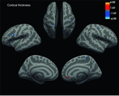
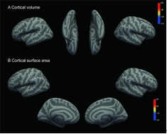
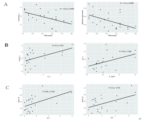
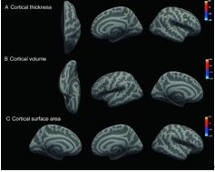
睡眠障碍是SARS-CoV-2感染康复者最常见的神经精神表现之一。既往一项纵向随访研究表明,从新冠肺炎中恢复的睡眠障碍患者的脑结构存在异常。然而,目前对新冠肺炎导致的睡眠障碍的神经影像学研究主要集中在长新冠中,尚缺乏急性感染期数据,这限制了对新冠肺炎导致的睡眠障碍的发病机制的了解。因此,这项纵向研究拟探索感染急性期患者的大脑结构是否发生变化,并通过3个月的随访观察其长期变化。研究共纳入26例患有睡眠障碍的新冠肺炎患者(51.5 ± 13.57岁,8女18男)、27例无睡眠障碍的新冠肺炎患者(47.33 ± 15.98岁,9女18男)和31名匹配的健康对照者(49.19 ± 17.51岁,9女22男),其中11例患有睡眠障碍的新冠肺炎患者在3个月进行纵向分析。结果发现,患有睡眠障碍的新冠肺炎患者几乎所有脑叶的结构都发生了变化,其中侧左额下回岛盖部和左侧楔前叶的皮质厚度与匹兹堡睡眠质量指数呈负相关。与健康对照组相比,新冠肺炎患者海马及其子区体积均发生了变化。3个月的纵向随访结果显示,与基线相比,患有睡眠障碍的新冠肺炎患者额顶叶皮质的大脑结构指数(皮质厚度、皮质灰质体积和皮质表面积)发生了变化。上述结果表明,新冠肺炎引起的睡眠障碍患者的脑结构异常始于感染急性期,且在3个月随访时持续存在。这填补了新冠急性期睡眠障碍患者神经影像证据的空白,有助于更好地理解新冠肺炎引起的睡眠障碍的病理机制。

https://orcid.org/0000-0002-7565-2607 (Demao Deng)

关键词: 睡眠障碍,  SARS-CoV-2,  新冠肺炎,  皮质厚度,  皮质灰质体积,  皮质表面积,  皮质下体积,  海马,  磁共振成像,  大脑结构

Abstract: Sleep disturbances are among the most prevalent neuropsychiatric symptoms in individuals who have recovered from severe acute respiratory syndrome coronavirus 2 infections. Previous studies have demonstrated abnormal brain structures in patients with sleep disturbances who have recovered from coronavirus disease 2019 (COVID-19). However, neuroimaging studies on sleep disturbances caused by COVID-19 are scarce, and existing studies have primarily focused on the long-term effects of the virus, with minimal acute phase data. As a result, little is known about the pathophysiology of sleep disturbances in the acute phase of COVID-19. To address this issue, we designed a longitudinal study to investigate whether alterations in brain structure occur during the acute phase of infection, and verified the results using 3-month follow-up data. A total of 26 COVID-19 patients with sleep disturbances (aged 51.5 ± 13.57 years, 8 women and 18 men), 27 COVID-19 patients without sleep disturbances (aged 47.33 ± 15.98 years, 9 women and 18 men), and 31 age- and gender-matched healthy controls (aged 49.19 ± 17.51 years, 9 women and 22 men) were included in this study. Eleven COVID-19 patients with sleep disturbances were included in a longitudinal analysis. We found that COVID-19 patients with sleep disturbances exhibited brain structural changes in almost all brain lobes. The cortical thicknesses of the left pars opercularis and left precuneus were significantly negatively correlated with Pittsburgh Sleep Quality Index scores. Additionally, we observed changes in the volume of the hippocampus and its subfield regions in COVID-19 patients compared with the healthy controls. The 3-month follow-up data revealed indices of altered cerebral structure (cortical thickness, cortical grey matter volume, and cortical surface area) in the frontal-parietal cortex compared with the baseline in COVID-19 patients with sleep disturbances. Our findings indicate that the sleep disturbances patients had altered morphology in the cortical and hippocampal structures during the acute phase of infection and persistent changes in cortical regions at 3 months post-infection. These data improve our understanding of the pathophysiology of sleep disturbances caused by COVID-19.

Key words: brain structure,  cortical gray matter volume,  cortical surface area,  cortical thickness,  hippocampus,  magnetic resonance imaging,   Pittsburgh Sleep Quality Index,  severe acute respiratory syndrome coronavirus 2 infections,  sleep disturbances,  sub-cortical volume