中国神经再生研究(英文版) ›› 2026, Vol. 21 ›› Issue (9): 4378-4389.doi: 10.4103/NRR.NRR-D-25-00338

• 原著:脑损伤修复保护与再生 • 上一篇    下一篇

 Hedgehog通路可逆转缺氧诱导人脑类器官的铁死亡


  • 出版日期:2026-09-15 发布日期:2026-05-22

Hedgehog signaling activation rescues hypoxia-induced ferroptosis in human brain organoids 

Simeng Yi1, 2, #, Min Huang1, 2, #, Chunmei Xian1, 2, Xi Kong1, 2, Shigang Yin1, 2, Jianhua Peng1, 2, 3, Hongda Li4, Yong Jiang1, 2, 3, *, Bingqing Xie1, 2, *, Huangfan Xie1, 2, *   

  1. 1Laboratory of Neurological Diseases and Brain Function, The Affiliated Hospital, Southwest Medical University, Luzhou, Sichuan Province, China; 
    2Institute of Brain Science, Southwest Medical University, Luzhou, Sichuan Province, China; 
    3Department of Neurosurgery, The Affiliated Hospital, Southwest Medical University, Luzhou, Sichuan Province, China; 
    4Institute for Brain Science and Disease, Key Laboratory of Major Brain Disease and Aging Research (Ministry of Education), Chongqing Medical University, Chongqing, China
  • Online:2026-09-15 Published:2026-05-22
  • Contact: Huangfan Xie, PhD, xie1991@swmu.edu.cn; Bingqing Xie, PhD, bingqingxie@swmu.edu.cn; Yong Jiang, MD, jiangyong@swmu.edu.cn.
  • Supported by:
    This work was supported by the National Natural Science Foundation of China, Nos. U24A20689 (to YJ), 32100454 (to HL); Sichuan Science and Technology Program, Nos. 2022YFS0615 (to BX), 2025ZNSFSC1019 (to HX); and Collaborative Fund of Southwest Medical University and Luzhou Government, No. 2024LZXNYDJ005 (to JP).

摘要:

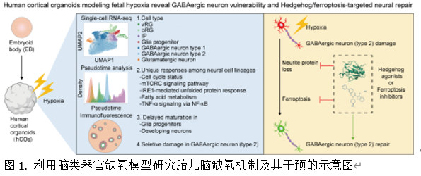
胎儿缺氧会扰乱神经发育,特别是正在发育的大脑对缺氧损伤极为敏感;然而,具体易受损的细胞类型及其潜在的分子机制仍未被充分研究。实验建立了一个人脑类器官模型,该模型能够再现妊娠早期至中期胎儿缺氧的病理生理过程。通过单细胞转录组学技术,在这些类器官中鉴定出7种神经细胞谱系,包括皮质前体细胞和神经元。进一步分析揭示了不同细胞类型在缺氧条件下对mTORC1信号通路、脂肪酸合成、未折叠蛋白反应和先天免疫反应的特定响应。在发育方面,谷氨酸能神经元和γ-氨基丁酸能神经元的成熟显著延迟,而前体细胞受影响较小。在功能方面,鉴定出两种对缺氧敏感性不同的GABA能神经元亚型:更成熟的类型2神经元对缺氧最为敏感,表现为铁死亡激活和神经突蛋白表达受损(如MAP2);而较不成熟的类型1神经元则表现出一定耐受性。机制研究表明,药理学激活 hedgehog 通路可抑制铁死亡并恢复 2 型 GABA 能神经元在缺氧条件下突触蛋白 MAP2 的表达。这些发现揭示了缺氧易感性的谱系特异性模式,并确立了 hedgehog 通路调节作为胎儿脑缺氧损伤中潜在靶向神经保护策略的可行性。


https://orcid.org/0000-0002-6342-4316 (Huangfan Xie); 

https://orcid.org/0000-0001-9433-1641 (Bingqing Xie); 

https://orcid.org/0000-0002-0490-3405 (Yong Jiang)

Abstract:

Fetal hypoxia disrupts neurodevelopment. In particular, the developing brain is extremely vulnerable to hypoxia injury; however, the specific vulnerable cell types and their underlying molecular mechanisms remain underexplored. In the present study, we established a human brain organoid model that reproduced the pathophysiological process of fetal hypoxia during early to mid-gestation. Through single-cell transcriptomic technology, we identified seven neural lineages in these organoids, including cortical progenitors and neurons. Further analysis revealed the specific responses to hypoxia among different types of cells regarding the mechanistic target of rapamycin complex 1 signaling pathway, fatty acid synthesis, the unfolded protein response, and the innate immune response. In terms of development, the maturation of glutamatergic and γ-aminobutyric acid-ergic neurons was significantly delayed after hypoxia exposure, whereas progenitor cells were less affected. In terms of function, we identified two subtypes of γ-aminobutyric acid-ergic neurons with different sensitivities to hypoxia. The more mature type 2 neurons were the most sensitive to hypoxia, which manifested as ferroptosis activation and impaired expression of neurite proteins (e.g., microtubule-associated protein 2). By contrast, the less mature type 1 neurons showed some tolerance to hypoxia. A mechanistic study revealed that pharmacological activation of the hedgehog pathway can inhibit ferroptosis and restore expression of the neurite protein microtubule-associated protein 2 in type 2 γ-aminobutyric acid-ergic neurons under hypoxia. Collectively, these findings delineate the lineage-specific patterns of hypoxia vulnerability and establish hedgehog pathway regulation as a potential target for neuroprotective strategies in fetal brain hypoxic injury.