周围神经损伤
-
Figure 7|Morphology and identification of primary Schwann cells.
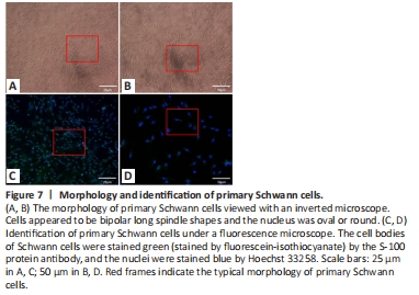
Schwann cells were successfully cultured by double enzyme digestion. The cells had a normal bipolar long spindle shape, the cells were slender, and the nucleus was oval or round. The immunohistochemical identification showed that the cell body of the Schwann cells was positive for S-100. The cultured primary Schwann cells had good purity and met the needs of subsequent experiments (Figure 7).
Figure 8|Screening of the drug concentration in primary Schwann cells.

Schwann cells were seeded in a biomimetic microfluidic chip, and the NGF complete medium (40 ng/mL) and bFGF-containing complete medium (10 ng) were pumped at a constant rate from Inlets 1 and 2 at a rate of 0.1 (μL/min). After 72 hours, the 22.86 ng/mL NGF + 4.29 ng/mL bFGF group had the best proliferation effect. Therefore, such a growth factor concentration combination strategy was selected for subsequent experiments (Figure 8).
Figure 15|Effects of NGF-bFGF-PLGA sustained-release microspheres on the myelinated nerve fibers in the sciatic nerve of rats with sciatic nerve injury (osmic acid staining).
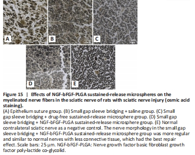
Twelve weeks after surgery, the distal nerve in each group was stained with osmic acid. The epithelium suture group had an irregular distribution of myelinated nerve fibers and more connective tissue, and the axons were different in size. Nerve regeneration was better in the small gap sleeve bridging group than in the epithelium suture group; however, it was still poorer than the normal nerve group. In the small gap sleeve bridging + NGF-bFGF-PLGA microsphere sustained-release group, the myelinated nerve fibers regenerated, and the nerve morphology was more regular, similar to normal nerves, and the connective tissue was less, which had better repair effect than the other groups (Figure 15).
Figure 17|Effects of NGF-bFGF-PLGA sustained-release microspheres on the gastrointestinal histological changes of rats with sciatic nerve injury (hematoxylin and eosin staining).
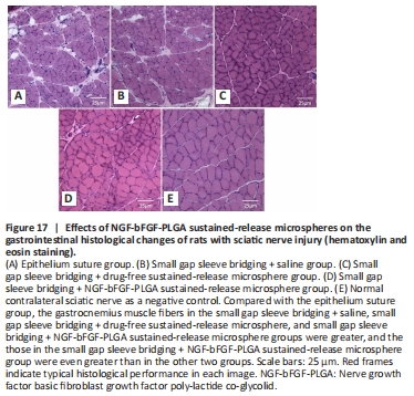
Twelve weeks after surgery, the muscles (gastrocnemius) were stained by hematoxylin and eosin. Our results showed that muscle fibers in the epithelium suture group were severely atrophied, and there was a large amount of collagen fiber growth. In the small gap sleeve bridging + saline, small gap sleeve bridging + drug-free sustained-release microsphere, and small gap sleeve bridging + NGF-bFGF-PLGA sustained-release microsphere groups, atrophy was milder, the gastrocnemius fiber was fuller, and the cut surface was neater. These findings suggest that small gap sleeve bridging + saline, small gap sleeve bridging + drug-free sustained-release microsphere, and small gap sleeve bridging + NGF-bFGF-PLGA sustained-release microspheres promote the functional recovery of peripheral nerve innervated muscle. The optimal repair effect was observed in the small gap sleeve bridging + NGF-bFGF-PLGA sustained-release microsphere group (Figure 17).