周围神经损伤
-
Figure 3|Effects of PRP on morphological recovery of regenerated nerves in rabbits with sciatic nerve injury.
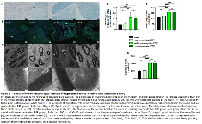
Toluidine blue staining was used to calculate the number of regenerated myelinated nerve fibers in the regions of nerve defects in rabbits. In the medium- and high-concentration PRP groups, there were more regenerated myelinated nerve fibers compared with those in the model and low-concentration PRP groups (P < 0.01). Immunofluorescent staining of NEFH was performed to assess the levels of neurofilament protein. The immunopositivity of neurofilaments was higher in the medium- and high-concentration PRP groups than in the model and low-concentration PRP groups (P < 0.001). Transmission electron microscopy was used to assess the thickness of the myelin sheath of myelinated nerve fibers, which indicates the maturity of the fibers. The number of regenerated nerve fibers in the medium- and high-concentration PRP groups was higher than that in the model and low-concentration PRP groups (P < 0.01; Figure 3A–G).
Figure 4|Effects of PRP on tricep recovery in rabbits with sciatic nerve injury.
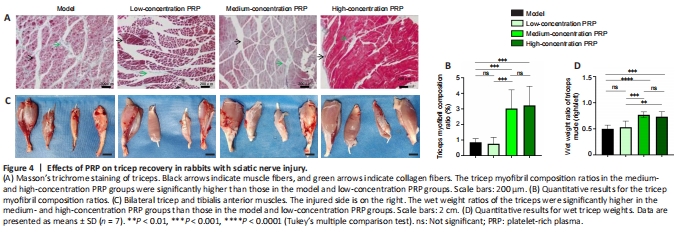
Masson’s trichrome staining of the triceps was performed to evaluate the tricep myofibril composition ratio (Figure 4A). The degree of muscle fibrosis in the medium- and high-concentration PRP groups was lower than that in the model and low-concentration PRP groups. Masson’s staining showed that the tricep myofibril composition ratio in the medium- and high-concentration PRP groups was significantly higher than that in the model and low-concentration PRP groups (P < 0.05). There was no significant difference in tricep myofibril composition ratio between the medium- and high-concentration PRP groups (P > 0.05) or between the low-concentration PRP and model groups (P > 0.05; Figure 4B). The wet weight ratio of the triceps was calculated to assess the extent of denervation of the triceps (Figure 4C). The wet weight ratio of the triceps was significantly higher in the medium- and high-concentration PRP groups than that in the model and low-concentration PRP groups (P < 0.01). There was no significant difference in the wet weight ratio of the triceps between the medium- and high-concentration PRP groups (P > 0.05) or between the model and low-concentration PRP groups (P > 0.05; Figure 4D).
点击此处查看全文