周围神经损伤
-
Figure 4|Effect of the granular hydrogel nerve guidance conduit on the FG-labeled motoneurons in the spinal anterior horn of rats with sciatic nerve injury 16 weeks after surgery.
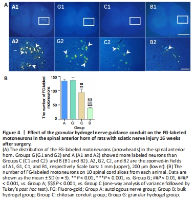
Figure 5|Effect of the granular hydrogel nerve guidance conduit on the FG-labeled sensory neurons in the DRG of rats with sciatic nerve injury 16 weeks after surgery.
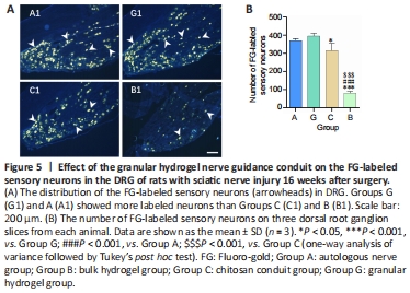
The numbers of motoneurons in the spinal anterior horn and sensory neurons in the DRG were examined with FG retrograde labeling. As shown in Figures 4 and 5, the granular hydrogel and autologous nerve groups showed no significant differences in the number of labeled motoneurons and sensory neurons (P = 0.9989 and P = 0.5625), while the granular hydrogel group showed more labeled neurons than the bulk hydrogel group (P < 0.0001 and P < 0.0001) and chitosan conduit group (P = 0.0015 and P = 0.0114).
Figure 6|Effect of the granular hydrogel nerve guidance conduit on the morphology analysis of regenerated nerves in rats with sciatic nerve injury 16 weeks after surgery.
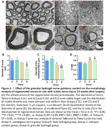
Myelinated nerve fibers in the sciatic nerve were examined with TEM 16 weeks after surgery. The diameters of the nerve fibers in the granular hydrogel and autologous nerve groups were visibly larger, and the distribution of myelin sheaths was more compact and uniform than the chitosan conduit and bulk hydrogel groups, as observed in Figure 6A. In Figure 6B, the diameter of the axons of the granular hydrogel group did not significantly differ from that of the autologous nerve group (P = 0.9896) or chitosan conduit group (P = 0.3928); however the granular hydrogel group was significantly larger than the bulk hydrogel group (P = 0.0034). The diameter of the myelinated fibers of the granular hydrogel group did not significantly differ from the autologous nerve group (P = 0.5234), and was significantly greater than the chitosan conduit group (P = 0.0126) and bulk hydrogel group (P = 0.0001; Figure 6C). The G-ratio value of the regenerated nerves of the granular hydrogel group did not significantly differ from the autologous nerve group (P = 0.3112); however, the granule hydrogel group was significantly less than the chitosan conduit group (P = 0.0042) and bulk hydrogel group (P = 0.0022; Figure 6D). These results indicated that the myelin sheath of the sciatic nerve in the granular hydrogel group was significantly thicker than that in the chitosan conduit and bulk hydrogel groups and was similar to that in the autologous nerve group.
Figure 7|Effect of the granular hydrogel nerve guidance conduit on the regenerated nerves in rats with sciatic nerve injury 16 weeks after the surgery.
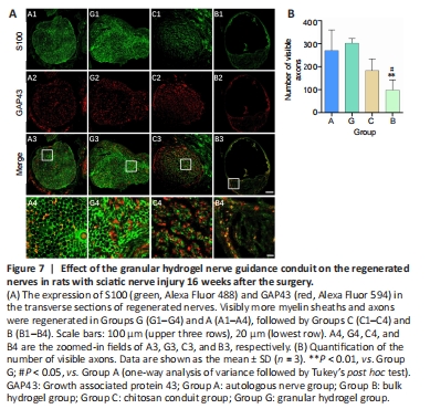
Sixteen weeks after surgery, the sciatic nerve was examined by immunochemical staining with anti-S100 and anti-GAP43. GAP43 is an axon membrane protein that is related to neuronal regeneration and growth (Chen et al., 2012). S100 protein is widely distributed in Schwann cells, astrocytes, and oligodendrocytes (Schlaepfer and Micko, 1978). Immunofluorescence histochemical staining of a normal sciatic nerve illustrating the positively labeled axons and myelin sheaths is shown in Additional Figure 2. As shown in Figure 7A, the regenerated myelin sheaths in the granular hydrogel and autologous nerve groups were relatively round or oval and the nerve fibers in the granular hydrogel group had a larger diameter than the chitosan conduit and bulk hydrogel groups. As shown in Figure 7B, the granular hydrogel group showed no significant difference in the number of GAP43-labeled axons from the autologous nerve group (P = 0.9013) and chitosan conduit group (P = 0.1211). The granular hydrogel group exhibited significantly more GAP43-labeled axons than the bulk hydrogel group (P = 0.0099). The results also clearly revealed a greater diameter of the regenerated nerve in the granular hydrogel group than those in the chitosan conduit and bulk hydrogel groups.
Figure 8|Effect of the granular hydrogel nerve guidance conduit on the gastrocnemius muscle atrophy in rats with sciatic nerve injury 16 weeks after surgery.
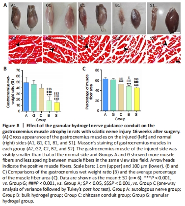
Results of the gastrocnemius muscle recovery 16 weeks after surgery are illustrated in Figure 8. The injured side (left) of the gastrocnemius muscle of each rat was obviously smaller than the normal side (right). As shown in Figure 8A, the gastrocnemius muscle of the surgical side was visibly smaller than that of the normal side and the granular hydrogel and autologous nerve groups had smaller gaps between the muscle fibers. The wet weight ratio of the granular hydrogel group showed no significant difference from that of the autologous nerve group (P = 0.9536) and chitosan conduit group (P = 0.4540); however, its wet weight ratio was significantly greater than that of the bulk hydrogel group (P < 0.0001) and silicone tube group (P < 0.0001). Furthermore, the area ratio of the gastrocnemius muscle fibers in the granular hydrogel group did not significantly differ from the autologous nerve group (P = 0.7917); however, its area ratio was significantly greater than the bulk hydrogel (P < 0.0001), chitosan conduit (P < 0.0001), and silicone tube groups (P < 0.0001; Figure 8B and C).