神经退行性病
-
Figure 1|The expression and the functionality of P-gp and the expression of Aβ in the brains of APP/PS1 mice compared with those in age-matched WT mice at different ages.
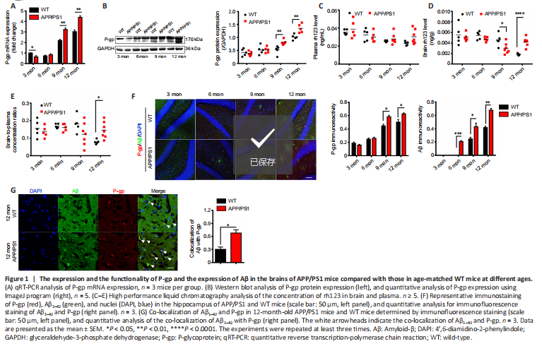
The expression of P-gp mRNA and protein in the brains of APP/PS1 mice and WT mice at different ages was measured by qRT-PCR and western blot assay. The expression of P-gp in both WT and APP/PS1 mice tended to increase with age, especially in APP/PS1 mice. At 3 months of age, the expression of P-gp in APP/PS1 mice was lower than that of WT mice, while at 6 months of age, the expression of P-gp in APP/PS1 was similar to that in WT mice; at 9 and 12 months of age, the expression of P-gp in APP/PS1 mice was significantly higher than that in WT mice (P < 0.01; Figure 1A and B).
P-gp functionality is represented by brain-to-plasma partition of the P-gp substrate rh123, and a decreased brain-to-plasma partition implicates increased P-gp functionality in brain. We measured brain and plasma concentrations of rh123 in APP/PS1 and WT mice at different ages, and the ratios of brain-to-plasma concentration were calculated. In the plasma, the concentrations of rh123 of both mice exhibited no evident difference at all test ages. In the brain, the concentrations of rh123 of both mice were equivalent at 3 and 6 months of age but different at 9 and 12 months of age (part of the data in Figure 1 were adopted from our previous report (Du and Yang, 2014)). At 3 and 6 months of age, the ratios of brain-to-plasma concentration of rh123 of the two mice were similar, suggesting that the functionalities of P-gp were not different. However, the functionality of P-gp in APP/PS1 mice was higher at 9 months of age and significantly lower at 12 months of age compared with that of WT mice (P < 0.05; Figure 1C–E).
The expression of P-gp of APP/PS1 mice tended to increase with age, with significantly higher expression at 9 and 12 months of age than that of WT mice (P < 0.01; Figure 1A and B). The functionality of P-gp of APP/PS1 mice did not change with age but was significantly lower than that of WT mice at 12 months of age (P < 0.05; Figure 1C–E). These results indicate that the expression of P-gp in APP/PS1 mice increased in the early stage of AD (9 months old (Sompol et al., 2021)) and the functionality of P-gp was impaired in the later stage of AD (12 months old (Wang et al., 2016)).
P-gp accumulates around deposited Aβ in the hippocampi of APP/PS1 mice
To study the relationship between P-gp and Aβ, double immunostaining was performed on brain samples of WT and APP/PS1 mice at all test ages. Aβ deposition began to appear in APP/PS1 mice at 6 months of age and in WT mice at 9 months of age. Over time, Aβ deposition increased; the deposition was significantly higher at 6, 9, and 12 months of age in APP/PS1 mice than in WT mice (P < 0.05; Figure 1F). Along with the increase of Aβ deposition, the P-gp expression also increased. The enrichment of P-gp surrounding Aβ deposition in the hippocampi of APP/PS1 mice was observed at 9 and 12 months of age, especially at the later age (Figure 1F). The co-localization of Aβ with P-gp was observed in the hippocampi of APP/PS1 mice at 12 months (Figure 1G). Taken together, these results suggest that the expression of P-gp in the brains of APP/PS1 mice is associated with the pathological progression of AD.
点击此处查看全文