脑损伤
-
Figure 2|General observations in the sham and intraventricular hemorrhage groups.
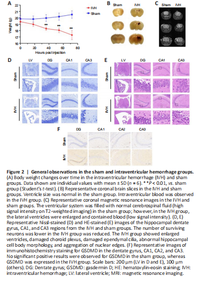
Body weight was lower in the IVH group than the sham group at different time points (Figure 2A), showing the damaging effect of IVH induction. Brain tissue sections and 7.0 T MRI showed enlargement of the lateral ventricle after IVH (Figure 2B and C). Nissl staining showed that the number of surviving neurons was lower in the IVH group than the sham group (Figure 2D). HE staining revealed ventricular enlargement, choroid plexus damage, destruction of ependymal cilia in the lateral ventricle region, and hippocampal cell body atrophy and nuclear edge aggregation in the IVH group (Figure 2E). Expression of GSDMD was higher in the IVH group than the sham group, demonstrating that IVH induced the activation of GSDMD and promoted pyroptosis (Figure 2F).
Figure 3| Expression of pyrolytic proteins was increased after intraventricular hemorrhage.

The expression of GSDMD, ASC, NLRP3, caspase-1, and IL-1β in the hippocampus after IVH was higher in the IVH group than the sham group; most proteins reached a peak at 72 hours (Figure 3A–F). Similarly, double immunofluorescence staining showed that GSDMD and NLRP3 co-localized with neurons. The percentage of GSDMD and NLRP3-positive neurons in the DG region was significantly higher in the IVH group than the sham group at 72 hours (P < 0.001) and 24 hours (P < 0.05) (Figure 3G–J). Based on our experimental results, 72 hours after IVH was identified as the time point for neuronal pyroptosis.
Figure 4| Effects of TUG on the expression of endoplasmic reticulum stress proteins after intraventricular hemorrhage.

Western blot results among the sham, IVH + TUG-891, IVH + DMOS, and IVH groups 72 hours after IVH showed that the expression of endoplasmic reticulum stress proteins was higher in the IVH groups and that expression was suppressed by TUG-891 (Figure 4A–E). Double immunofluorescence staining showed that the percentages of ATF-4- and CHOP-positive neurons in the DG region were higher in the IVH groups than the sham group (P < 0.001) and that TUG-891 attenuated these changes (P < 0.05 for CHOP and P < 0.01 for ATF-4) (Figure 4F–I). These findings demonstrate the effective inhibition of endoplasmic reticulum stress by TUG-891.
Figure 5|The inhibitory effects of TUG on pyroptosis after intraventricular hemorrhage.

Western blot results among the sham, IVH + TUG-891, IVH + DMSO, and IVH groups 72 hours after IVH showed that the expression of the pyroptosis proteins GSDMD, ACS, NLPR3, caspase-1, and IL-1β was suppressed by TUG-891 (Figure 5A–F). Double immunofluorescence staining showed that the percentages of NLRP3- (P < 0.001) and GSDMD-positive neurons (P < 0.001) was lower in mice pretreated with TUG-891, confirming that TUG-891 inhibits neuronal pyroptosis (Figure 5G–J).