神经退行性病
-
Figure 1| Effect of MPP+ on PHB2 protein expression and mitochondrial function in SH-SY5Y cells.
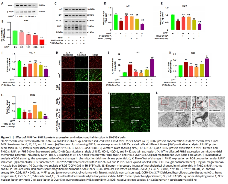
Human neuroblastoma SH-SY5Y cells are a dopaminergic neuronal cell line used as an in vitro model for neurotoxicity experiments. MPP+ inhibits the activity of oxidative respiratory chain complex I in mitochondria, which can cause oxidative stress injury in dopaminergic neurons and affect neuronal mitophagy (Ramsay et al., 1986). We treated SH-SY5Y cells with 1 mM MPP+, and found decreased PHB2 expression at 12 hours (Figure 1A and B). Next, we determined whether PHB2 affected the intracellular antioxidative response and mitochondrial function. Nrf2 is a nuclear factor erythroid 2-related factor that can translocate to the nucleus and recognize the enhancer sequence of antioxidant response elements. In turn, this increases expression of HO-1 and NQO-1 to resist various stress injuries (Hayes and Dinkova-Kostova, 2014). In our study, we found MPP+ significantly reduced the expression of Nrf2, HO-1, and NQO-1. Moreover, overexpression of PHB2 increased protein expression of Nrf2, HO-1, and NQO-1, while silencing of PHB2 decreased levels of these anti-oxidative stress proteins under MPP+ treatment (Figure 1C–G). We also examined the effect of PHB2 on JC-1 fluorescence (Figure 1H and I) and ROS accummulation (Figure 1J and K). We found that overexpression of PHB2 increased the mitochondrial membrane potential and reduced ROS levels in the presence of MPP+. Silencing PHB2 decreased the mitochondrial membrane potential and increased ROS levels. Finally, we observed the mitochondrial state of PHB2-shRNA-treated cells by electron microscopy (Figure 1L). Normal morphology of the mitochondrial inner ridge was observed in the control group. PHB2-shRNA treatment changed the morphology of the mitochondrial inner ridge, while MPP+ treatment caused significant swelling of the inner ridge.
Figure 2| Effect of PHB2 on mitophagy in a PD cell model.
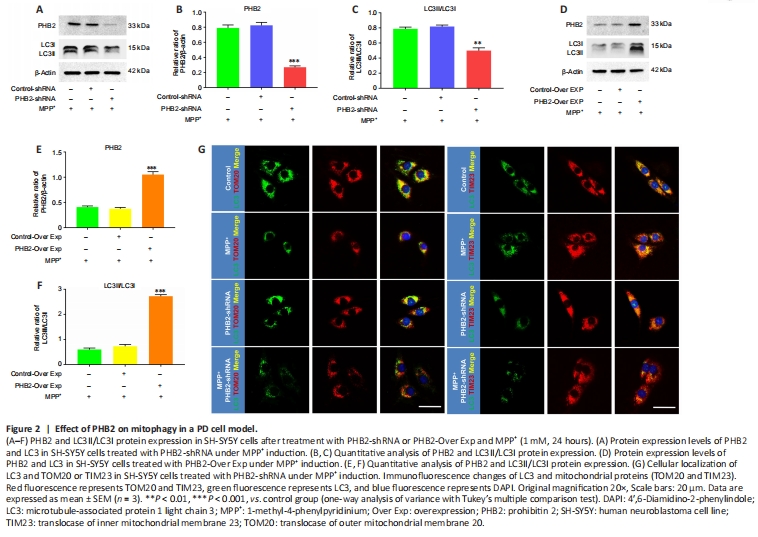
As PHB2 promotes mitophagy by recruiting LC3II (Wei et al., 2017), we further examined whether PHB2-mediated mitophagy is involved in PD. Changes in the LC3II/LC3I ratio were observed in MPP+-treated SH-SY5Y cells by silencing or overexpression of PHB2. The LC3II/LC3I ratio was lower after PHB2 silencing than in the control group (Figure 2A–C). The LC3II/LC3I ratio was higher following overexpression of PHB2 than in the control group (Figure 2D–F). We used the mitochondrial outer membrane protein, TOM20, and the mitochondrial inner membrane protein, TIM23, as mitochondrial localization markers. Silencing of PHB2 decreased colocalization of LC3 and TOM20 and TIM23 (Figure 2G).
Figure 3| Parkin increases PHB2/LC3-mediated mitophagy in SH-SY5Y cells.
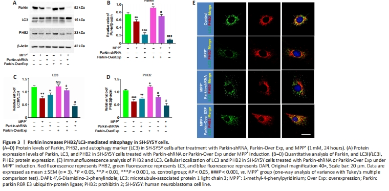
Next, we investigated whether Parkin regulation affected protein expression and interaction of PHB2 and LC3. We found that Parkin overexpression increased protein expression of PHB2 and the LC3II/LC3I ratio. Silencing of Parkin decreased the LC3II/LC3I ratio and protein expression of PHB2 under MPP+ treatment (Figure 3A–D). Immunofluorescence staining showed that silencing of Parkin decreased co-localization of PHB2 and LC3 under MPP+ treatment, whereas overexpression of Parkin increased co-localization (Figure 3E). Taken together, these results suggest that Parkin affects mitophagy by regulating the expression and binding of PHB2 to LC3.
Figure 6|Silencing PHB2 inhibits mitophagy and aggravates dopaminergic neuronal loss in PD mice.
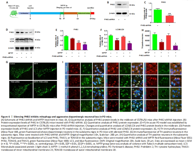
To observe the role of PHB2 in PD, we injected AAV9 PHB2-shRNA into the striatum of PD mice (Figure 6B and C). We found that silencing PHB2 decreased the LC3II/LC3I ratio (Figure 6D–F). TH immunofluorescence of substantia nigra neurons indicated that MPTP significantly reduced the number of TH-positive neurons, and silencing of PHB2 aggravated MPTP-induced loss of dopaminergic neurons (Figure 6G and H). Moreover, silencing of PHB2 under MPTP treatment reduced co-localization of LC3 with PHB2, TIM23, and TOM20 (Figure 6I).