脑损伤
-
Figure 4|Cav3.2 KO suppresses oxidative stress in the hippocampus after cerebral ischemia/reperfusion injury.
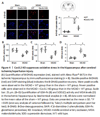
To explore the effect of Cav3.2 on CIRI-induced oxidative stress, we detected 8-OHdG expression in the hippocampus after CIRI. Compared with the WT + MCAO group, the Cav3.2 KO + MCAO group had reduced 8-OHdG levels in the hippocampus (Figure 4A). Similarly, the Cav3.2 KO + MCAO group had improved GSH-Px and SOD activities, and decreased MDA levels in the hippocampus when compared with those in the WT + MCAO group (Figure 4B–D). These results suggest that Cav3.2 KO suppresses oxidative stress in the hippocampus after CIRI.
Figure 6|Cav3.2 KO attenuates cell apoptosis after cerebral ischemia/reperfusion injury.
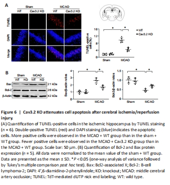
Next, we evaluated the effect of Cav3.2 KO on CIRI-induced cell apoptosis. Cav3.2 KO markedly reduced cell apoptosis in the ischemic hippocampal tissue in the MCAO mice (Figure 6A). Moreover, Cav3.2 KO remarkably downregulated Bax expression and upregulated Bcl-2 expression after MCAO (Figure 6B). These results suggest that Cav3.2 KO reduces cell apoptosis after CIRI.