中国神经再生研究(英文版) ›› 2024, Vol. 19 ›› Issue (11): 2480-2487.doi: 10.4103/1673-5374.390966

• 原著:脑损伤修复保护与再生 • 上一篇    下一篇

Cav3.2通道调控脑缺血再灌注损伤:有前途的干预靶点


  • 出版日期:2024-11-15 发布日期:2024-03-29
  • 基金资助:
    安徽省自然科学基金优青项目(2208085Y32),安徽省教育厅科学研究杰青项目(2022AH020076),湖北省陈孝平科技发展基金项目(CXPJJH12000005-07-115)

Cav3.2 channel regulates cerebral ischemia/reperfusion injury: a promising target for intervention

Feibiao Dai1, 2, 3, #, Chengyun Hu1, 2, 3, #, Xue Li1, 2, 3, #, Zhetao Zhang3, 4, Hongtao Wang2, 3, Wanjun Zhou2, 3, Jiawu Wang2, 3, Qingtian Geng2, 3, *, Yongfei Dong3, 5, *, Chaoliang Tang2, 3, *   

  1. 1Graduate School, Wannan Medical College, Wuhu, Anhui Province, China; 2Department of Anesthesiology, The First Affiliated Hospital of USTC, Division of Life Sciences and Medicine, University of Science and Technology of China, Hefei, Anhui Province, China; 3Core Facility Center for Medical Sciences, The First Affiliated Hospital of USTC (Anhui Provincial Hospital), Hefei, Anhui Province, China; 4Department of Pharmacy, The First Affiliated Hospital of USTC, Division of Life Sciences and Medicine, University of Science and Technology of China, Hefei, Anhui Province, China; 5Department of Neurosurgery, The First Affiliated Hospital of USTC, Division of Life Sciences and Medicine, University of Science and Technology of China, Hefei, Anhui Province, China
  • Online:2024-11-15 Published:2024-03-29
  • Contact: Chaoliang Tang, MD, PhD, chaolt@ustc.edu.cn; Yongfei Dong, MD, dyf.w@163.com; Qingtian Geng, MD, gengqingtian@sina.com.
  • Supported by:
    This study was supported by the Natural Science Foundation of Anhui Province of China, No. 2208085Y32; Scientific Research Plan Project of Anhui Province of China, No. 2022AH020076; and the Chen Xiao-Ping Foundation for the Development of Science and Technology of Hubei Province, No. CXPJJH12000005-07-115 (all to CT).

摘要:

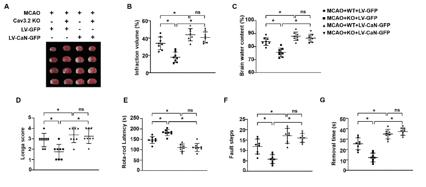
在脑缺血再灌注损伤中,钙流入大脑神经元引发神经元死亡,且多种钙通道参与了脑缺血再灌注损伤。Cav3.2通道是T型钙通道的主要亚型,有研究发现T型钙通道阻滞剂匹莫齐德和米贝拉地尔可预防脑缺血再灌注损伤诱导的脑损伤,但是Cav3.2通道在脑缺血再灌注损伤中的作用尚未阐明。为此,实验分别以大脑中动脉阻塞诱导小鼠脑缺血再灌注损伤以及高糖缺氧/复氧诱导原代海马神经元以构建体内外脑缺血再灌注损伤模型,发现损伤的海马组织和原代海马神经元中Cav3.2表达显著上调。实验进一步建立Cav3.2基因敲除小鼠脑缺血再灌注损伤模型,发现Cav3.2敲除可显著减轻脑缺血再灌注损伤所致脑梗死体积、脑含水量以及神经功能障碍。此外,Cav3.2敲除还减弱了脑缺血再灌注损伤诱导的氧化应激、炎症反应和神经元凋亡。而在Cav3.2敲除小鼠海马中过表达钙调神经磷酸酶可抵消Cav3.2敲除对脑缺血再灌注损伤的保护作用。上述结果表明,Cav3.2敲除介导的神经保护功能是由钙调神经磷酸酶/激活T细胞核因子3信号传导介导的。这项研究表明,Cav3.2可能是脑缺血再灌注损伤的一个有前途的干预靶点。

https://orcid.org/0000-0002-6873-7821 (Qingtian Geng); https://orcid.org/0000-0002-1740-1375 (Yongfei Dong); 
https://orcid.org/0000-0002-1936-028X (Chaoliang Tang)

关键词: Cav3.2通道, 脑缺血再灌注, 脑卒中, 缺氧/复氧, 氧化应激, 炎症反应, 钙调神经磷酸酶, 激活T细胞核因子3, 海马, 原代海马神经元

Abstract: Calcium influx into neurons triggers neuronal death during cerebral ischemia/reperfusion injury. Various calcium channels are involved in cerebral ischemia/reperfusion injury. Cav3.2 channel is a main subtype of T-type calcium channels. T-type calcium channel blockers, such as pimozide and mibefradil, have been shown to prevent cerebral ischemia/reperfusion injury-induced brain injury. However, the role of Cav3.2 channels in cerebral ischemia/reperfusion injury remains unclear. Here, in vitro and in vivo models of cerebral ischemia/reperfusion injury were established using middle cerebral artery occlusion in mice and high glucose hypoxia/reoxygenation exposure in primary hippocampal neurons. The results showed that Cav3.2 expression was significantly upregulated in injured hippocampal tissue and primary hippocampal neurons. We further established a Cav3.2 gene-knockout mouse model of cerebral ischemia/reperfusion injury. Cav3.2 knockout markedly reduced infarct volume and brain water content, and alleviated neurological dysfunction after cerebral ischemia/reperfusion injury. Additionally, Cav3.2 knockout attenuated cerebral ischemia/reperfusion injury-induced oxidative stress, inflammatory response, and neuronal apoptosis. In the hippocampus of Cav3.2-knockout mice, calcineurin overexpression offset the beneficial effect of Cav3.2 knockout after cerebral ischemia/reperfusion injury. These findings suggest that the neuroprotective function of Cav3.2 knockout is mediated by calcineurin/nuclear factor of activated T cells 3 signaling. Findings from this study suggest that Cav3.2 could be a promising target for treatment of cerebral ischemia/reperfusion injury.

Key words: calcineurin, Cav3.2 channel, cerebral ischemia/reperfusion, hippocampus, hypoxia/reoxygenation, inflammatory response, nuclear factor of activated T cells 3, oxidative stress, primary hippocampal neurons, stroke