神经损伤与修复
-
Figure 2|Electric stimulation promotes photoreceptor survival in human retinal explant cultures.
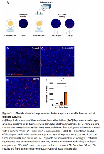
We next compared the neuroprotective potential of different ES waveforms in human post-mortem retinal explant cultures. Previous studies suggested that employing ES at a ramp waveform (20 Hz) or a series of rectangular pulse trains at increasing frequencies from 20–200 Hz improved retinal cell survival in vitro and in vivo (Enayati et al., 2020; Yu et al., 2020). It prompted us to evaluate the effects of these ES conditions on photoreceptor cell survival in culture. Using post-mortem human retinal tissue explants. The graphical summary of the experiment is presented in Figure 2A. Sham-stimulated explants served as the controls. Following 48 hours of incubation after ES, the survival of photoreceptors was assessed using rhodopsin immunostaining. In control cultures, substantial loss of the photoreceptors was observed, with only a few rhodopsin+ cells detectable (Figure 2B). Rectangular ES improved the number of surviving photoreceptors (Figure 2C), but this number did not reach significance (P > 0.05). Remarkably, the stimulation with ramp waveform resulted in dramatically increased photoreceptor survival after 48 hours of culture (Figure 2D). Quantitative analysis of the rhodopsin-positive cells in the explants (Figure 2E) indicated that ramp waveforms have a significant increase in the number of rhodopsin-positive surviving photoreceptors when compared with control (P < 0.05) or rectangular waveform (P < 0.05). These results suggest the potent neuroprotective effects of ramp waveform ES.
Figure 4|tcES increases the survival of photoreceptors.
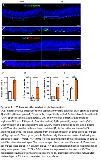
We counted photoreceptor cells in retinal sections to determine if tcES improves retinal and visual functions by preserving photoreceptors. Immunostaining revealed that tcES ameliorated the loss of B-opsins (Figure 4A) and Red/Green-opsins cells (Figure 4B). Quantitative analysis of B-opsins positive cells (P < 0.01; Figure 4C); Red/Green-opsins positive cells (P < 0.05; Figure 4D) and total opsin positive cells numbers (P < 0.05; Figure 4E) demonstrated significantly improved survival of B-opsin, R/G-opsin, and total Opsin-positive cells in the tcES group compared to sham-treated contralateral eyes). ONL thickness was also significantly increased (P < 0.001) in tcES-treated retinas compared to sham-treated eyes. This indicates improved survival of all types of photoreceptor cells after tcES treatment (Figure 4F). These results indicate tcES ameliorated photoreceptor loss in Rho–/– mice retina.
Figure 5|tcES increases bipolar cell sprouting.
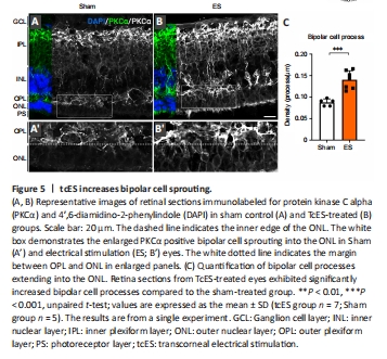
Bipolar cells have been shown to undergo structural changes and neurite sprouting that contribute to retinal function and recovery (Lewis et al., 1998). To evaluate if tcES affected bipolar cell plasticity, we performed immunolabeling for the bipolar cell-specific marker Protein kinase Cα (PKCα). We observed prominent upregulation of PKCα immunolabeling in the tcES-treated retina, primarily associated with bipolar cell processes, compared to the sham-treated retina (Figure 5A and B). We also noted an increase in bipolar cell processes that grew into the ONL. Quantitative analysis of the density of the bipolar cell dendrite extending into the ONL confirmed that tcES significantly increased the density of bipolar cell sprouting (P < 0.001) into the ONL compared to the sham-treated group (Figure 5C). Increased retinal plasticity and bipolar cell sprouting in response to the tcES may be an additional mechanism contributing to increased photoreceptor survival and function in the Rho–/– mice.