脊髓损伤
-
Figure 3 | NRMS reduces neuronal damage in the injured area of SCI rats.
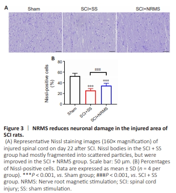
Next, we determined whether NRMS had effects on the pathological changes of the injured spinal cord. Nissl staining (Figure 3A) showed that the Nissl bodies in the Sham group were large and abundant. The majority of Nissl bodies in the SCI + SS group were small, some of which had already fragmented into scattered particles and exhibited numerous pathological changes with vacuolar structures. Compared with the SCI rats with sham stimulation, the NRMS-treated rats had a significant increase in the percentage of Nissl-positive cells (P < 0.001; Figure 3B) and less neuronal damage.
Figure 4 | NRMS enhances the structural plasticity of dendrites in the damaged spinal cord.
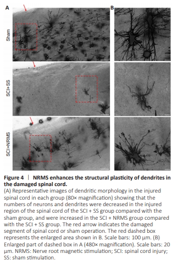
We then evaluated the effects of NRMS on neuronal dendrite morphology in the damaged spinal cord. We performed qualitative observations of neuronal dendrites 3 weeks after SCI, and found the injured region of the spinal cord of the SCI + SS group had formed glial scars and appeared to have a decrease in the number of neurons and dendrites compared with those of the Sham group (Figure 4A and B). Compared with the sham-stimulated SCI group, the SCI + NRMS group