中国神经再生研究(英文版) ›› 2024, Vol. 19 ›› Issue (7): 1559-1567.doi: 10.4103/1673-5374.387972

• 原著:脊髓损伤修复保护与再生 • 上一篇    下一篇

基于小鼠下肢深层肌肉运动神经元可视化三维空间分布的“信使区”假说


  • 出版日期:2024-07-15 发布日期:2023-11-28
  • 基金资助:
    国家自然科学基金面上项目(82072162)

A “messenger zone hypothesis” based on the visual three-dimensional spatial distribution of motoneurons innervating deep limb muscles

Chen Huang1, 2, #, Shen Wang1, 2, #, Jin Deng1, 2, Xinyi Gu1, 2, Shuhang Guo1, 2, Xiaofeng Yin1, 2, *   

  1. 1MoE Key Laboratory for Trauma Treatment and Nerve Regeneration, Peking University, Beijing, China; 2Department of Orthopedics and Trauma, Peking University People’s Hospital, Beijing, China
  • Online:2024-07-15 Published:2023-11-28
  • Contact: Xiaofeng Yin, PhD, xiaofengyin@bjmu.edu.cn.
  • Supported by:
    This study was continuously supported by the Chinese National General Program of the National Natural Science Foundation of China, No. 82072162 (to XY).

摘要:

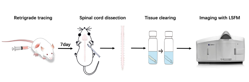
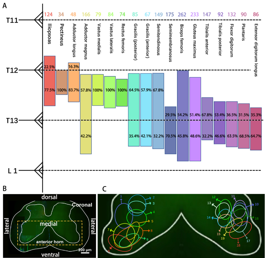
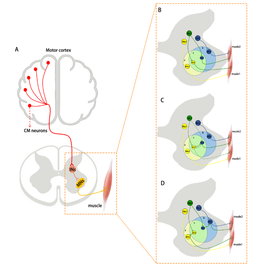
骨骼肌的协调收缩依赖于自身与多种脊髓运动神经元之间的选择性连接。然而,目前对支配不同肌肉的脊髓运动神经元的空间分布研究是极为有限的。此次实验结合与溶剂清除器官三维成像兼容(3DISCO)的多重逆行示踪方法,并利用光片荧光显微镜三维成像技术研究了股神经、闭孔神经、臀下神经、腓深神经以及胫神经支配的小鼠后肢深层肌肉不同运动神经元的空间分布和相对位置。此外,实验还提出了“信使区”假说,即在支配协同或拮抗剂肌群的运动神经元池之间存在交错区域(被定义为信使区),其中交错分布的神经元可能作为信使神经元参与肌肉协调。这项研究不仅揭示了支配小鼠不同深层肌肉的多个运动神经元池之间精确的相互位置关系,还更新和补充了支配小鼠肌肉的脊髓运动神经元的系统化三维视觉图谱,而且对于理解协调肌肉活动的潜在机制和运动回路的结构也具有积极意义。

https://orcid.org/0000-0001-9932-642X (Xiaofeng Yin)

关键词: 三维成像, 空间分布, 骨骼肌, 运动神经元, 组织光学清除, 多次逆行示踪, 肌肉协调

Abstract: Coordinated contraction of skeletal muscles relies on selective connections between the muscles and multiple classes of the spinal motoneurons. However, current research on the spatial location of the spinal motoneurons innervating different muscles is limited. In this study, we investigated the spatial distribution and relative position of different motoneurons that control the deep muscles of the mouse hindlimbs, which were innervated by the obturator nerve, femoral nerve, inferior gluteal nerve, deep peroneal nerve, and tibial nerve. Locations were visualized by combining a multiplex retrograde tracking technique compatible with three-dimensional imaging of solvent-cleared organs (3DISCO) and 3-D imaging technology based on lightsheet fluorescence microscopy (LSFM). Additionally, we propose the hypothesis that “messenger zones” exist as interlaced areas between the motoneuron pools that dominate the synergistic or antagonist muscle groups. We hypothesize that these interlaced neurons may participate in muscle coordination as messenger neurons. Analysis revealed the precise mutual positional relationships among the many motoneurons that innervate different deep muscles of the mouse. Not only do these findings update and supplement our knowledge regarding the overall spatial layout of spinal motoneurons that control mouse limb muscles, but they also provide insights into the mechanisms through which muscle activity is coordinated and the architecture of motor circuits.

Key words: 3-D imaging, motoneurons, multiple retrograde tracing, muscle coordination, skeletal muscle, spatial distribution, optical tissue clearing