中国神经再生研究(英文版) ›› 2025, Vol. 20 ›› Issue (8): 2408-2419.doi: 10.4103/NRR.NRR-D-23-01301

• 原著:视神经损伤修复保护与再生 • 上一篇    下一篇

AAV2-PDE6B 抑制细胞凋亡拯救Rd10 模型小鼠视网膜色素变性


  • 出版日期:2025-08-15 发布日期:2024-12-14

AAV2-PDE6B restores retinal structure and function in the retinal degeneration 10 mouse model of retinitis pigmentosa by promoting phototransduction and inhibiting apoptosis

Ruiqi Qiu1, 2, #, Mingzhu Yang1, 2, #, Xiuxiu Jin1, 2, 3, Jingyang Liu1, 2, Weiping Wang1, 2, Xiaoli Zhang1, 4, Jinfeng Han1, 4, Bo Lei1, 2, 3, 4, *   

  1. 1 Henan Eye Hospital, Henan Provincial People’s Hospital, People’s Hospital of Zhengzhou University, Zhengzhou, Henan Province, China; 2 Eye Institute, Henan Academy of Innovations in Medical Science, Zhengzhou, Henan Province, China; 3 Branch of National Clinical Research Center for Ocular Disease, Henan Provincial People’s Hospital, Zhengzhou, Henan Province, China; 4 Academy of Medical Science, Zhengzhou University, Zhengzhou, Henan Province, China
  • Online:2025-08-15 Published:2024-12-14
  • Contact: Bo Lei, MD, PhD, FARVO, bolei99@126.com.
  • Supported by:
    This study was supported by the National Natural Science Foundation of China, Nos. 82071008 (to BL) and 82004001 (to XJ); Medical Science and Technology Program of Health Commission of Henan Province, No. LHGJ20210072 (to RQ); and Science and Technology Department of Henan Province, No. 212102310307 (to XJ).

摘要:

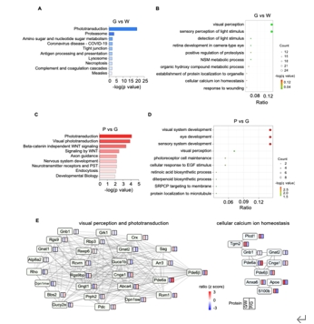
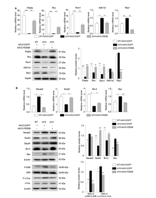
AAV是一种安全有效的基因运载工具,其介导的基因治疗已成为遗传性视网膜疾病的治疗的主流方向。动物实验证实AAV介导的PDE6B治疗能显著改善视网膜的结构与功能,然而AAV-PDE6B载体发挥治疗作用的具体分子机制以及分子水平上的变化尚不明确。为了解AAV2-PDE6B 改善视网膜色素变性视网膜功能的潜在分子机制。实验于rd10小鼠视网膜下注射AAV2-PDE6B后,使用暗适应和光适应视网膜电图、光学相干断层扫描和免疫荧光评估了对视网膜功能和结构的治疗效果。实验进一步进行了基于数据独立采集-质谱的蛋白质组分析,以研究蛋白质表达和通路富集,并通过实时 PCR 和 Western blot进一步筛选和验证。发现注射AAV2-PDE6B能显著提高PDE6β的水平,保护rd10小鼠的视网膜电图和核外层厚度。野生型小鼠和rd10小鼠的差异表达蛋白(DEPs)与视觉感知密切相关,AAV2-PDE6B治疗后视觉感知得到恢复。注射AAV2-PDE6B后,KEGG富集的最明显变化途径是光传导。此外,与光传导相关的蛋白 Pde6α,Rom1,Rho,Aldh1a1,Rbp1 表达也发生了逆转。rd10小鼠经AAV2-PDE6B处理后,Bax/Bcl-2,p-ERK/ERK和p-c-Fos/c-Fos水平均下降。实验数据表明,AAV2-PDE6B介导的基因治疗通过促进光传导和抑制 ERK1/2 介导的细胞凋亡发挥保护视网膜色素变性视网膜作用。

https://orcid.org/0000-0002-5497-0905 (Bo Lei)

Abstract: Retinitis pigmentosa is a group of inherited diseases that lead to retinal degeneration and photoreceptor cell death. However, there is no effective treatment for retinitis pigmentosa caused by PDE6B mutation. Adeno-associated virus (AAV)-mediated gene therapy is a promising strategy for treating retinitis pigmentosa. The aim of this study was to explore the molecular mechanisms by which AAV2- PDE6B rescues retinal function. To do this, we injected retinal degeneration 10 (rd10) mice subretinally with AAV2-PDE6B and assessed the therapeutic effects on retinal function and structure using dark- and light-adapted electroretinogram, optical coherence tomography, and immunofluorescence. Data-independent acquisition-mass spectrometry-based proteomic analysis was conducted to investigate protein expression levels and pathway enrichment, and the results from this analysis were verified by real-time polymerase chain reaction and western blotting. AAV2-PDE6B injection significantly upregulated PDE6β expression, preserved electroretinogram responses, and preserved outer nuclear layer thickness in rd10 mice. Differentially expressed proteins between wild-type and rd10 mice were closely related to visual perception, and treating rd10 mice with AAV2-PDE6B restored differentially expressed protein expression to levels similar to those seen in wild-type mice. Kyoto Encyclopedia of Genes and Genome analysis showed that the differentially expressed proteins whose expression was most significantly altered by AAV2-PDE6B injection were enriched in phototransduction pathways. Furthermore, the phototransductionrelated proteins Pde6α, Rom1, Rho, Aldh1a1, and Rbp1 exhibited opposite expression patterns in rd10 mice with or without AAV2-PDE6B treatment. Finally, Bax/Bcl-2, p-ERK/ERK, and p-c-Fos/c-Fos expression levels decreased in rd10 mice following AAV2-PDE6B treatment. Our data suggest that AAV2-PDE6B-mediated gene therapy promotes phototransduction and inhibits apoptosis by inhibiting the ERK signaling pathway and upregulating Bcl-2/Bax expression in retinitis pigmentosa.

Key words: apoptosis,  AAV2-PDE6B,  ERK1/2,  gene therapy,  phototransduction,  proteomics,  rd10,  retinitis pigmentosa