中国神经再生研究(英文版) ›› 2023, Vol. 18 ›› Issue (10): 2285-2290.doi: 10.4103/1673-5374.369124

• 原著:脑损伤修复保护与再生 • 上一篇    下一篇

由Notch信号通路介导的星形胶质细胞-神经元通讯:关注谷氨酸转运和突触可塑性


  • 出版日期:2023-10-15 发布日期:2023-03-29
  • 基金资助:
    国家自然科学基金;国家青年科学基金;盛京医院优秀科学基金和盛京医院345人才支持项目

Astrocyte-neuron communication mediated by the Notch signaling pathway: focusing on glutamate transport and synaptic plasticity

Ke-Xin Li, Meng Lu, Meng-Xu Cui, Xiao-Ming Wang*, Yang Zheng*   

  1. Department of Radiology, Shengjing Hospital of China Medical University, Shenyang, Liaoning Province, China
  • Online:2023-10-15 Published:2023-03-29
  • Contact: Yang Zheng, MD, zhengyang19871114@163.com; Xiao-Ming Wang, MD, wangxm024@163.com.
  • Supported by:
    This study was supported by the National Natural Science Foundation of China, Nos. 81871408 and 81271631 (to XMW), National Science Foundation for Young Scientists of China, No. 81801658 (to YZ), Outstanding Scientific Fund of Shengjing Hospital, No. 201402 (to XMW), and 345 Talent Support Project of Shengjing Hospital, No. 30B (to YZ).

摘要:

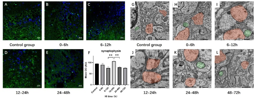
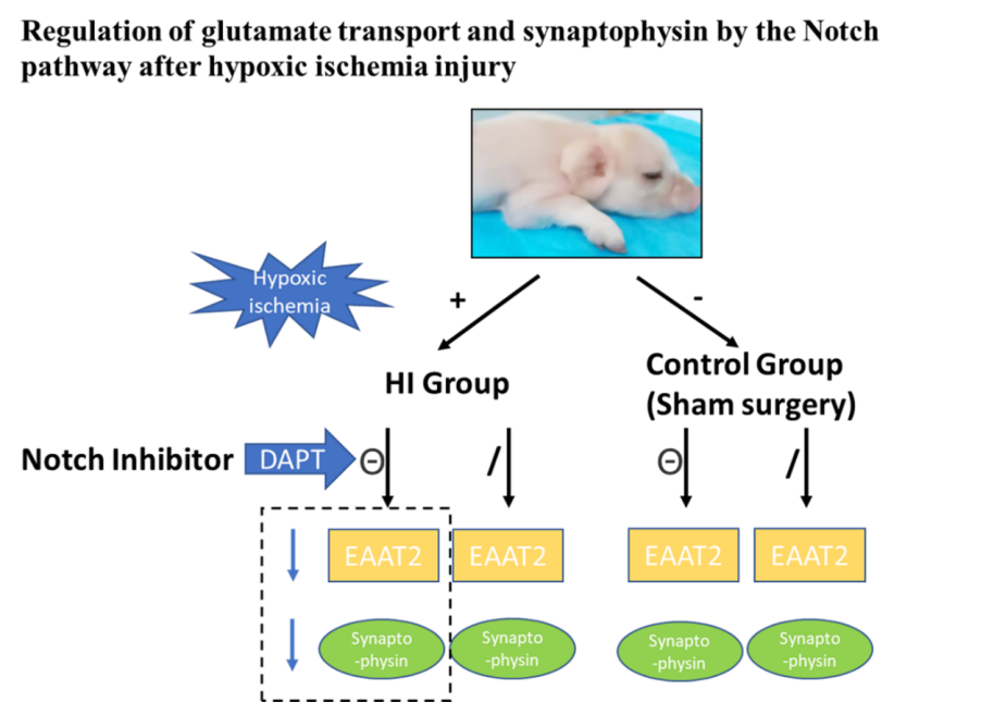
维持神经递质的平衡是星形胶质细胞对神经元的支持作用之一。其中,谷氨酸的平衡是维持突触功能和神经细胞活动的一个重要方面。然而,研究谷氨酸的动态变化、谷氨酸转运的分子机制以及对突触的影响尚需进一步研究。为探讨Notch信号通路介导的谷氨酸转运和突触可塑性的调节机制,实验以暂时性阻断双侧颈总动脉40min,同时机械吸入6%的氧气的方法来制作新生猪急性缺氧缺血性脑病模型。建模后,随机将动物分为Notch通路抑制组及Notch通路非抑制组,Notch通路抑制组动物以3.33 mg/mL的浓度腹腔注射Notch通路抑制剂(DAPT),非抑制组动物注射等剂量DMSO。应用免疫组化、免疫荧光及免疫印迹实验评估脑内蛋白表达情况。应用透射电镜观察脑内微观结构变化。在缺氧缺血性脑损伤早期(损伤后6-12h),谷氨酸转运体兴奋性氨基酸转运体2和突触蛋白下调,突触小泡数量减少,突触肿胀;在损伤后12-24h,Notch通路被激活,谷氨酸转运体兴奋性氨基酸转运体2和突触蛋白的表达增加,突触小泡数量略有增加。反过来,在应用Notch途径抑制剂后,谷氨酸转运体和突触蛋白的表达下降。这表明,缺氧缺血性脑损伤后星形胶质细胞-神经元的谷氨酸转运受Notch途径调节,并通过突触蛋白的表达影响囊泡释放和突触可塑性。

https://orcid.org/0000-0002-7452-0725 (Yang Zheng); https://orcid.org/ 0000-0003-0276-2466 (Xiao-Ming Wang)

Abstract: Maintaining glutamate homeostasis after hypoxic ischemia is important for synaptic function and neural cell activity, and regulation of glutamate transport between astrocyte and neuron is one of the important modalities for reducing glutamate accumulation. However, further research is needed to investigate the dynamic changes in and molecular mechanisms of glutamate transport and the effects of glutamate transport on synapses. The aim of this study was to investigate the regulatory mechanisms underlying Notch pathway mediation of glutamate transport and synaptic plasticity. In this study, Yorkshire neonatal pigs (male, age 3 days, weight 1.0–1.5 kg, n = 48) were randomly divided into control (sham surgery group) and five hypoxic ischemia subgroups, according to different recovery time, which were then further subdivided into subgroups treated with dimethyl sulfoxide or a Notch pathway inhibitor (N-[N-(3, 5-difluorophenacetyl-l-alanyl)]-S-phenylglycine t-butyl ester). Once the model was established, immunohistochemistry, immunofluorescence staining, and western blot analyses of Notch pathway-related proteins, synaptophysin, and glutamate transporter were performed. Moreover, synapse microstructure was observed by transmission electron microscopy. At the early stage (6–12 hours after hypoxic ischemia) of hypoxic ischemic injury, expression of glutamate transporter excitatory amino acid transporter-2 and synaptophysin was downregulated, the number of synaptic vesicles was reduced, and synaptic swelling was observed; at 12–24 hours after hypoxic ischemia, the Notch pathway was activated, excitatory amino acid transporter-2 and synaptophysin expression was increased, and the number of synaptic vesicles was slightly increased. Excitatory amino acid transporter-2 and synaptophysin expression decreased after treatment with the Notch pathway inhibitor. This suggests that glutamate transport in astrocytes-neurons after hypoxic ischemic injury is regulated by the Notch pathway and affects vesicle release and synaptic plasticity through the expression of synaptophysin.

Key words: astrocyte, astrocyte-neuron communication, glutamate, glutamate transporter, hypoxic-ischemic injury, magnetic resonance spectroscopy, neonate, Notch signaling pathway, plasticity, synapse