中国神经再生研究(英文版) ›› 2021, Vol. 21 ›› Issue (5): 2031-2039.doi: 10.4103/NRR.NRR-D-24-00431

• 原著:脊髓损伤修复保护与再生 • 上一篇    下一篇

磁共振成像示踪超顺磁性纳米铁剂标记间充质基质细胞修复脊髓损伤的能力


  • 出版日期:2026-05-15 发布日期:2025-08-23
  • 基金资助:
    国家重点研发计划(2017YF0104302,2017YF0104304)

Magnetic resonance imaging tracing of superparamagnetic iron oxide nanoparticle–labeled mesenchymal stromal cells for repairing spinal cord injury

Xiaoli Mai1, #, Yuanyuan Xie2, #, Zhichong Wu3, #, Junting Zou1 , Jiacheng Du4 , Yunpeng Shen5 , Hao Liu6 , Bo Chen7 , Mengxia Zhu1 , Jiong Shi8 , Yang Chen5 , Bing Zhang1, *, Zezhang Zhu3, *, Bin Wang2, *, Ning Gu9   

  1. 1 Department of Radiology, Nanjing Drum Tower Hospital, Affiliated Hospital of Medical School, Nanjing University, Nanjing, Jiangsu Province, China;  2 Clinical Stem Cell Center, Nanjing Drum Tower Hospital, Affiliated Hospital of Medical School, Nanjing University, Nanjing, Jiangsu Province, China;  3 Department of Spinal Surgery, Nanjing Drum Tower Hospital, Affiliated Hospital of Medical School, Nanjing University, Nanjing, Jiangsu Province, China;  4 State Key Laboratory of Bioelectronics and Jiangsu Key Laboratory of Biomaterials and Devices, School of Biological Sciences and Medical Engineering, Southeast University, Nanjing, Jiangsu Province, China;  5 Laboratory of Image Science and Technology, Southeast University, Nanjing, Jiangsu Province, China;  6 Department of Neurosurgery, Nanjing Drum Tower Hospital, Affiliated Hospital of Medical School, Nanjing University, Nanjing, Jiangsu Province, China;  7 Institute of Materials Science and Devices, School of Materials Science and Engineering, Suzhou University of Science and Technology, Suzhou, Jiangsu Province, China;  8 Department of Pathology, Nanjing Drum Tower Hospital, Affiliated Hospital of Medical School, Nanjing University, Nanjing, Jiangsu Province, China;  9 Nanjing Key Laboratory for Cardiovascular Information and Health Engineering Medicine, Institute of Clinical Medicine, Nanjing Drum Tower Hospital, Affiliated Hospital of Medical School, Nanjing University, Nanjing, Jiangsu Province, China
  • Online:2026-05-15 Published:2025-08-23
  • Contact: Bin Wang, MD, PhD, wanbin022800@126.com; Bing Zhang, MD, PhD, zhangbing_nj@vip.163.com; Zezhang Zhu, MD, PhD, zhuzezhang@126.com.
  • Supported by:
    This study was supported by the National Key R&D Program of China, Nos. 2017YFA0104302 (to NG and XM) and 2017YFA0104304 (to BW and ZZ).

摘要:

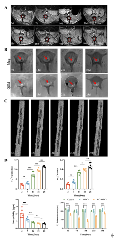
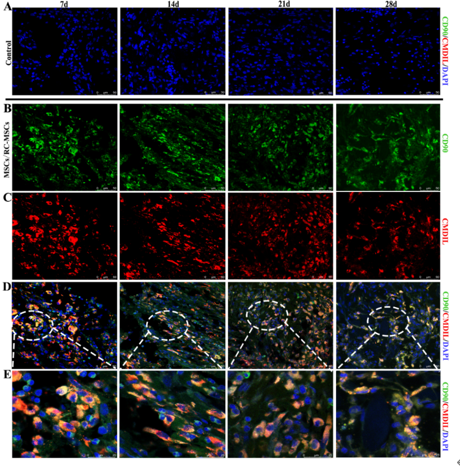
间充质基质细胞移植是治疗多种全身性和弥漫性疾病的一种有效而有前景的方法。但是目前尚不明确移植间充质基质细胞在体内的生物学特性,包括细胞活性、分布、迁移和在人体内的转归。传统的细胞示踪方法无法用于临床。使用超顺磁性氧化铁纳米粒子作为造影剂可在磁共振成像下对移植细胞进行观察。2016年国家医药产品监督管理局已批准一种新型超顺磁性氧化铁纳米粒子瑞存可用于临床试验的造影剂。此次实验使用比格犬制作半横断急性脊髓损伤模型,而后移植瑞存标记间充质基质细胞进行治疗。结果显示,瑞存标记的间充质基质细胞也能有效修复受损脊髓纤维,并部分恢复急性脊髓损伤动物的神经功能。且T2*WI上可见损伤脊髓两侧存在低信号区域。UTE-QSM量化结果显示,瑞存标记的间充质基质细胞可在损伤脊髓中稳定存在超过4周。提示磁共振成像在有效追踪瑞存标记的间充质基质细胞迁移变化及评估脊髓损伤修复能力方面有潜力。

https://orcid.org/0000-0003-3981-8849 (Bin Wang)
关键词: 间充质基质细胞, 神经功能, 神经元再生, 急性脊髓损伤, 动态迁移, 超顺磁氧化铁纳米粒子, 瑞存, 弥散张量成像, 定量易感图, 可修复性

Abstract: Mesenchymal stromal cell transplantation is an effective and promising approach for treating various systemic and diffuse diseases. However, the biological characteristics of transplanted mesenchymal stromal cells in humans remain unclear, including cell viability, distribution, migration, and fate. Conventional cell tracing methods cannot be used in the clinic. The use of superparamagnetic iron oxide nanoparticles as contrast agents allows for the observation of transplanted cells using magnetic resonance imaging. In 2016, the National Medical Products Administration of China approved a new superparamagnetic iron oxide nanoparticle, Ruicun, for use as a contrast agent in clinical trials. In the present study, an acute hemi-transection spinal cord injury model was established in beagle dogs. The injury was then treated by transplantation of Ruicun-labeled mesenchymal stromal cells. The results indicated that Ruicunlabeled mesenchymal stromal cells repaired damaged spinal cord fibers and partially restored neurological function in animals with acute spinal cord injury. T2*-weighted imaging revealed low signal areas on both sides of the injured spinal cord. The results of quantitative susceptibility mapping with ultrashort echo time sequences indicated that Ruicun-labeled mesenchymal stromal cells persisted stably within the injured spinal cord for over 4 weeks. These findings suggest that magnetic resonance imaging has the potential to effectively track the migration of Ruicun-labeled mesenchymal stromal cells and assess their ability to repair spinal cord injury.

Key words: acute spinal cord injury, diffusion tensor imaging, dynamic migration, mesenchymal stromal cells, neural function, neuronal regeneration, quantitative susceptibility mapping, repairability, ruicun, superparamagnetic iron oxide nanoparticle