中国神经再生研究(英文版) ›› 2023, Vol. 18 ›› Issue (10): 2307-2314.doi: 10.4103/1673-5374.369122

• 原著:视神经损伤修复保护与再生 • 上一篇    下一篇

抑制Srgap2可减轻小鼠视网膜神经节细胞变性


  • 出版日期:2023-10-15 发布日期:2023-03-29
  • 基金资助:
    国家自然科学基金项目(81770918,31871383);浙江省自然科学基金项目(LY16H120006);温州医科大学部门基金项目(89214018)

Srgap2 suppression ameliorates retinal ganglion cell degeneration in mice

Yi-Jing Gan1, 2, Ying Cao1, Zu-Hui Zhang1, Jing Zhang1, Gang Chen1, Ling-Qin Dong1, Tong Li1, Mei-Xiao Shen1, Jia Qu1, *, Zai-Long Chi1, *   

  1. 1State Key Laboratory of Ophthalmology, Optometry and Vision Science; Eye Hospital, Wenzhou Medical University, Wenzhou, Zhejiang Province, China; 2Department of Ophthalmology, The First Affiliated Hospital of University of Science and Technology of China, Division of Life Sciences and Medicine, University of Science and Technology of China, Hefei, Anhui Province, China
  • Online:2023-10-15 Published:2023-03-29
  • Contact: Zai-Long Chi, MD, PhD, zailong.chi@eye.ac.cn; Jia Qu, MD, PhD, jia.qu@eye.ac.cn.
  • Supported by:
    This study was supported by the National Natural Science Foundation of China, Nos. 81770918 (to ZLC), 31871383 (to TL); the Natural Science Foundation of Zhejiang Province, No. LY16H120006 (to ZLC); and the Departmental Funds from Wenzhou Medical University, No. 89214018 (to ZLC).

摘要:

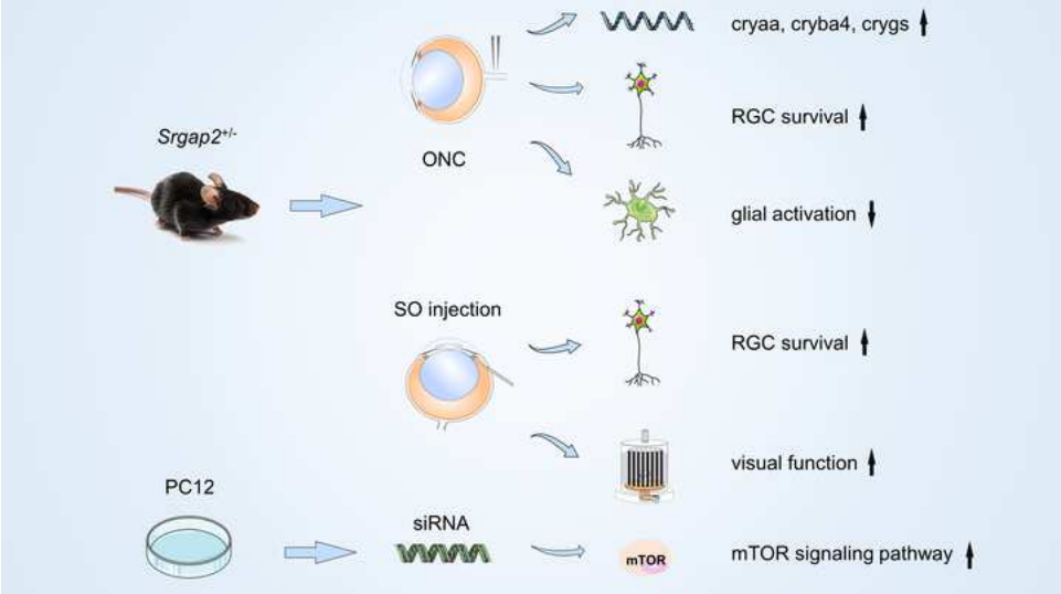
Slit-Robo-Rho-GTPase激活蛋白2(SRGAP2)在轴突引导、神经元迁移、突触形成和神经再生中发挥着重要的作用,但其在神经视网膜变性疾病中的作用尚不清楚。实验首先发现,正常小鼠视网膜在胚胎期即开始表达SRGAP2蛋白,且主要位于成熟的视网膜神经节细胞层和内核层,而视神经夹闭损伤后,小鼠视网膜和视神经中SRGAP2蛋白水平升高。进一步以Srgap2杂合子(Srgap2+/-)小鼠建立视神经夹闭模型,发现抑制Srgap2可提高视网膜神经节细胞的存活率,降低眼内压,抑制胶质细胞活化,并部分恢复视网膜功能。而体外实验中沉默Srgap2基因则能促进PC12细胞中哺乳动物雷帕霉素靶蛋白信号通路的激活。视网膜转录组测序发现,Srgap2+/-小鼠视网膜中与视神经损伤保护相关的小热休克蛋白基因(cryaa,cryba4和crygs)水平上调。上述结果表明抑制Srgap2基因表达可通过减缓胶质细胞的过度活化,并激活神经保护相关的哺乳动物雷帕霉素靶蛋白通路和小热休克蛋白的表达,改善视网膜神经节细胞变性和挽救部分视神经功能。

https://orcid.org/0000-0002-3617-6425 (Zai-Long Chi); https://orcid.org/0000-0001-5729-567X (Jia Qu)

关键词: Srgap2, 视网膜神经节细胞变性, 视神经夹闭, 硅油, 眼高压, 星形胶质细胞, 晶体蛋白, 哺乳动物雷帕霉素靶蛋白, 视神经病变, 神经保护

Abstract: Slit-Robo GTPase-activating protein 2 (SRGAP2) plays important roles in axon guidance, neuronal migration, synapse formation, and nerve regeneration. However, the role of SRGAP2 in neuroretinal degenerative disease remains unclear. In this study, we found that SRGAP2 protein was first expressed in the retina of normal mice at the embryonic stage and was mainly located in the mature retinal ganglion cell layer and the inner nuclear layer. SRGAP2 protein in the retina and optic nerve increased after optic nerve crush. Then, we established a heterozygous knockout (Srgap2+/–) mouse model of optic nerve crush and found that Srgap2 suppression increased retinal ganglion cell survival, lowered intraocular pressure, inhibited glial cell activation, and partially restored retinal function. In vitro experiments showed that Srgap2 suppression activated the mammalian target of rapamycin signaling pathway. RNA sequencing results showed that the expression of small heat shock protein genes (Cryaa, Cryba4, and Crygs) related to optic nerve injury were upregulated in the retina of Srgap2+/– mice. These results suggest that Srgap2 suppression reduced the robust activation of glial cells, activated the mammalian target of rapamycin signaling pathway related to nerve protein, increased the expression of small heat shock protein genes, inhibited the degeneration of retinal ganglion cells, and partially restored optic nerve function. 

Key words: astrocyte, crystallins, intraocular hypertension, microglia, mTOR, neuroprotection, optic nerve crush, optic neuropathy, RGC degeneration,  , Srgap2