中国神经再生研究(英文版) ›› 2024, Vol. 19 ›› Issue (5): 1112-1118.doi: 10.4103/1673-5374.385310

• 原著:视神经损伤修复保护与再生 • 上一篇    下一篇

抑制酪蛋白激酶2可促进急性眼压升高后视网膜神经节细胞存活


  • 出版日期:2024-05-15 发布日期:2023-10-31
  • 基金资助:
    国家自然科学基金项目(81570849,81100931);广东省自然科学基金项目(2015A0303134462020A01515011413)

Casein kinase-2 inhibition promotes retinal ganglion cell survival after acute intraocular pressure elevation

Meng Wang1, 2, Shi-Qi Yao1, 2, Yao Huang3, Jia-Jian Liang1, Yanxuan Xu1, Shaowan Chen1, Yuhang Wang1, Tsz Kin Ng1, 2, 4, #br# Wai Kit Chu4, Qi Cui1, 4, Ling-Ping Cen1, 2, *#br#   

  1. 1Joint Shantou International Eye Center of Shantou University and The Chinese University of Hong Kong, Shantou, Guangdong Province, China; 2Shantou University Medical College, Shantou, Guangdong Province, China; 3Beijing Tongren Eye Center, Beijing Tongren Hospital, Capital Medical University, Beijing, China; 4Department of Ophthalmology and Visual Sciences, The Chinese University of Hong Kong, Hong Kong Special Administrative Region, China
  • Online:2024-05-15 Published:2023-10-31
  • Contact: Ling-Ping Cen, MD, PhD, cenlp@hotmail.com.
  • Supported by:
    This study was supported by the National Natural Science Foundation of China, Nos. 81570849, 81100931; the Natural Science Foundation of Guangdong Province of China, Nos. 2015A030313446, 2020A1515011413 (all to LPC).

摘要:

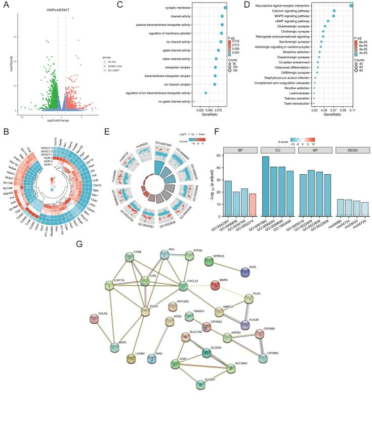
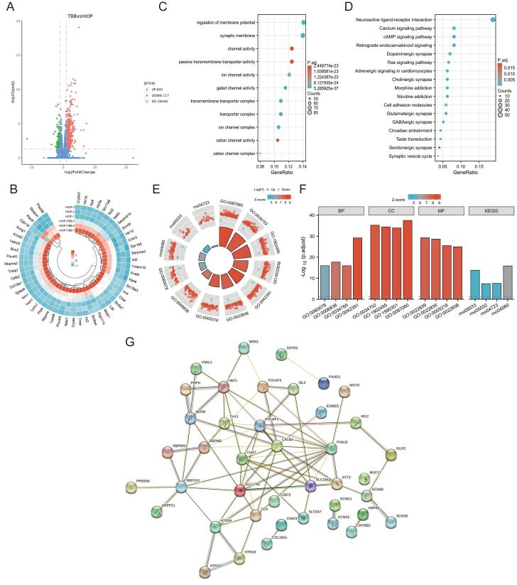
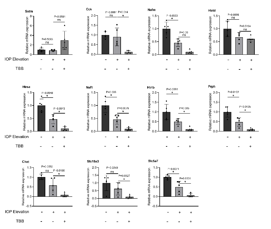
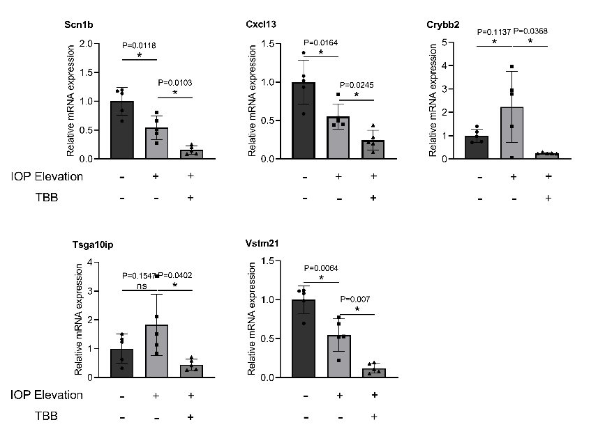
眼压升高可引起视网膜神经节细胞死亡,这是青光眼临床可逆危险因素之一,且青光眼是不可逆失明的主要原因。作者团队既往研究已发现,酪蛋白激酶2抑制可促进视神经损伤后大鼠视网膜神经节细胞的存活和轴突再生。为了解这一作用的机制,实验首先将成年大鼠眼压升高至75mmHg并持续2h,然后经玻璃体注射酪蛋白激酶2抑制剂2-二甲基氨基-4,5,6,7-四溴苯并咪唑(TBB)和2-二甲基氨基-4,5,6,7-四溴苯并咪唑(DMAT)进行治疗。结果可见,TBB和DMAT抑制酪蛋白激酶2显著促进了视网膜神经节细胞活性,同时减少了浸润巨噬细胞的数量。进一步转录组学分析显示,MAPK信号通路参与眼压升高,但与酪蛋白激酶2抑制无关。此外,酪蛋白激酶2抑制可下调参与眼压升高的基因(Cck, Htrsa, Nef1, Htrlb, Prph, Chat, Slc18a3, Slc5a7, Scn1b, Crybb2, Tsga10ip以及Vstm21)的表达。由此表明,抑制酪蛋白激酶2可通过抑制巨噬细胞激活来提高眼压升高后大鼠视网膜神经节细胞活性。

https://orcid.org/0000-0003-3876-0606 (Ling-Ping Cen)

关键词: 青光眼, 高眼压, 视网膜神经节细胞, 酪蛋白激酶2, 巨噬细胞

Abstract: Intraocular pressure elevation can induce retinal ganglion cell death and is a clinically reversible risk factor for glaucoma, the leading cause of irreversible blindness. We previously demonstrated that casein kinase-2 inhibition can promote retinal ganglion cell survival and axonal regeneration in rats after optic nerve injury. To investigate the underlying mechanism, in the current study we increased the intraocular pressure of adult rats to 75 mmHg for 2 hours and then administered a casein kinase-2 inhibitor (4,5,6,7-tetrabromo-2-azabenzimidazole or 2-dimethylamino-4,5,6,7-tetrabromo-1H-benzimidazole) by intravitreal injection. We found that intravitreal injection of 4,5,6,7-tetrabromo-2-azabenzimidazole or 2-dimethylamino-4,5,6,7-tetrabromo-1H-benzimidazole promoted retinal ganglion cell survival and reduced the number of infiltrating macrophages. Transcriptomic analysis showed that the mitogen activated protein kinase signaling pathway was involved in the response to intraocular pressure elevation but was not modulated by the casein kinase-2 inhibitors. Furthermore, casein kinase-2 inhibition downregulated the expression of genes (Cck, Htrsa, Nef1, Htrlb, Prph, Chat, Slc18a3, Slc5a7, Scn1b, Crybb2, Tsga10ip, and Vstm21) involved in intraocular pressure elevation. Our data indicate that inhibition of casein kinase-2 can enhance retinal ganglion cell survival in rats after acute intraocular pressure elevation via macrophage inactivation. 

Key words: casein kinase-2, glaucoma, intraocular pressure elevation, macrophages, retinal ganglion cells