中国神经再生研究(英文版) ›› 2026, Vol. 21 ›› Issue (10): 4965-4977.doi: 10.4103/NRR.NRR-D-25-01263

• 原著:脊髓损伤修复保护与再生 • 上一篇    下一篇

M1巨噬细胞分泌CXCL12驱动脊髓损伤后神经源性异位骨化#br#
#br#


  • 出版日期:2026-10-15 发布日期:2026-06-15

M1 macrophage-derived CXCL12 drives neurogenic heterotopic ossification following spinal cord injury

Yulei Xie1, 2, #, Yaomin Luo1, #, Xin Chen1, Yinxu Wang1, Wei Song3, *, Hua Ling3, *   

  1. 1Department of Rehabilitation Medicine, Affiliated Hospital of North Sichuan Medical College, Nanchong, Sichuan Province, China; 
    2School of Rehabilitation, Capital Medical University, Beijing, China; 
    3Department of Rehabilitation Engineering, China Rehabilitation Science Institute, Beijing, China

  • Online:2026-10-15 Published:2026-06-15
  • Contact: Wei Song, MS, wsongabc@163.com; Hua Ling, PhD, walter0615@163.com.
  • Supported by:
    This study was supported by the Fundamental Research Funds for Central Public Welfare Research Institutes (China Rehabilitation Science Institute), No. 2023CZ-10 (to WS) and the Research Innovation Team of North Sichuan Medical College, No. CBYTD-2025A05 (to YX).

摘要:

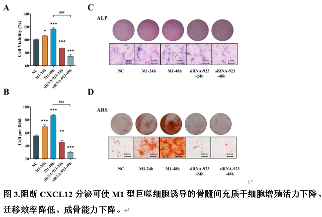
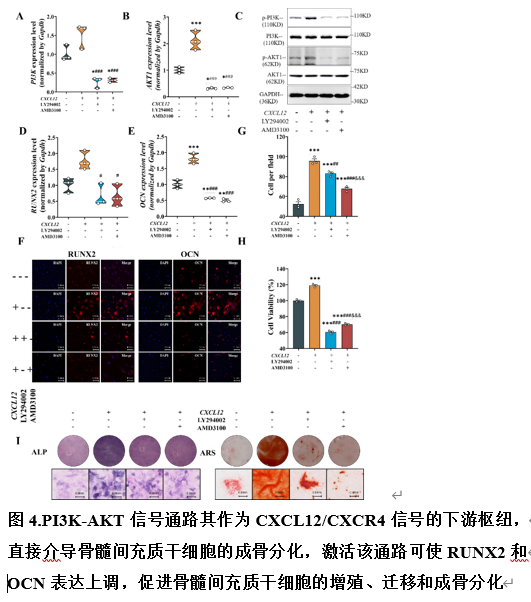
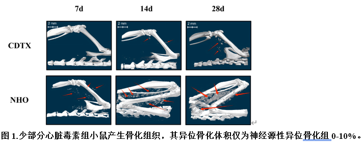
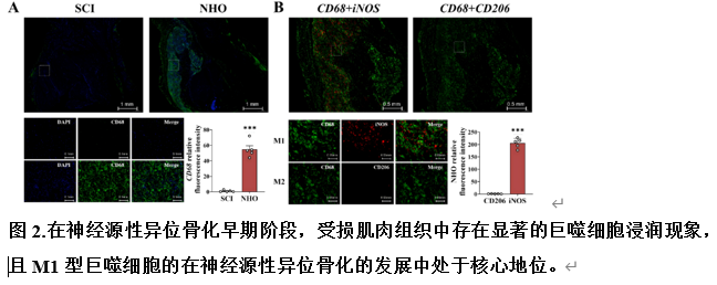
脊髓损伤患者常发生神经源性异位骨化,其发病机制尚不完全清楚。现有的研究模型难以准确模拟复杂的病理过程。为建立可靠的神经源性异位骨化研究模型,阐明其发病机制,探索早期干预策略,此次实验成功构建了脊髓损伤诱导的神经源型异位骨化小鼠模型。结果可见明显的异位骨形成、髋关节和膝关节活动受限以及运动功能障碍,并伴有成骨标志物ALP、RUNX2、SOX9和OCN的表达升高。蛋白质组学和定量聚合酶链反应分析显示,在神经源性异位骨化组中,CXCL12、CXCR4和LYN上调,而CXCL1下调。体内实验证实,M1巨噬细胞在早期异位骨组织周围的肌肉中异常积聚,CXCL12表达显著升高。体外研究进一步揭示,M1巨噬细胞是CXCL12分泌的主要来源,其细胞培养上清液促进骨髓间充质干细胞的增殖、迁移和成骨分化潜力。CXCL12通过与CXCR4受体结合激活PI3K/AKT通路,从而驱动骨髓间充质干细胞的成骨分化。这些发现表明,脊髓损伤后神经源性异位骨化的病理过程与CXCL12-CXCR4-PI3K-AKT信号轴的激活之间存在因果关系,该信号轴通过M1巨噬细胞极化调节骨髓间充质干细胞功能。实验揭示了驱动骨髓间充质干细胞成骨分化的关键机制,为脊髓损伤后神经源性异位骨化的早期预警和靶向治疗提供了新的方向。


https://orcid.org/0009-0000-5045-7082 (Wei Song); https://orcid.org/0000-0002-3219-5039 (Hua Ling)
关键词: CXCL12, CXCR4, 异位骨化, 巨噬细胞, 迁移, 矿化, 成骨, PI3K/AKT, 增殖, 脊髓损伤

Abstract: Patients with spinal cord injury frequently develop neurogenic heterotopic ossification, whose pathogenesis remains incompletely understood. Existing research models struggle to accurately simulate the complex pathological process. To establish a reliable neurogenic heterotopic ossification research model, elucidate its pathogenesis, and explore early intervention strategies, this study successfully developed a spinal cord injury-induced neurogenic heterotopic ossification mouse model. Significant ectopic bone formation, restricted hip and knee joint mobility, and motor dysfunction were observed, accompanied by elevated expression of the osteogenic markers alkaline phosphatase, runt-related transcription factor 2, sex-determining region Y-box 9, and osteocalcin . Proteomics and quantitative polymerase chain reaction analysis revealed upregulation of chemokine (C-X-C motif) ligand (CXCL)12, C-X-C chemokine receptor type 4 (CXCR4), and LYN proto-oncogene, whereas CXCL1 was downregulated in the neurogenic heterotopic ossification group. In vivo experiments confirmed abnormal accumulation of M1 macrophages in muscles surrounding early ectopic bone tissue, with markedly elevated CXCL12 expression. In vitro studies further revealed that M1 macrophages are the primary source of CXCL12 secretion, and their cell culture supernatants promote the proliferation, migration, and osteoblastic differentiation potential of bone marrow mesenchymal stem cells. Mechanistically, CXCL12 activates the phosphatidylinositol 3-kinase/protein kinase B pathway by binding to the CXCR4 receptor, thereby driving the osteogenic differentiation of bone marrow mesenchymal stem cells. These findings indicate a causal relationship between the pathological process of neurogenic heterotopic ossification following spinal cord injury and the activation of the CXCL12-CXCR4-phosphatidylinositol 3-kinase-protein kinase B signaling axis, which modulates bone marrow mesenchymal stem cell function through M1 macrophage polarization. This study reveals a key mechanism driving the osteogenic differentiation of bone marrow mesenchymal stem cells, providing new directions for early warning and targeted treatment of neurogenic heterotopic ossification following spinal cord injury.

Key words: CXCL12, CXCR4, heterotopic ossification, macrophage, migration, mineralization, osteogenic, PI3K/AKT, proliferation, spinal cord injury