中国神经再生研究(英文版) ›› 2026, Vol. 21 ›› Issue (8): 3831-3841.doi: 10.4103/NRR.NRR-D-25-00006

• 原著:退行性病与再生 • 上一篇    下一篇

多模态磁共振成像与RNA测序共同揭示9月龄3xTg-AD小鼠大脑病理学特征


  • 出版日期:2026-08-18 发布日期:2026-04-28

Multimodal MRI combined with RNA sequencing reveals pathological signatures in the 9-month-old 3×Tg-AD mouse brain

Yongxin Li1, 2, *, #, Ziling Tang1, #, Maohua Yao1, Yun Ran1, Zuocheng Qiu1, 2, *   

  1. 1Guangzhou Key Laboratory of Formula-pattern Research Center, School of Traditional Chinese Medicine, Jinan University, Guangzhou, Guangdong Province, China; 
    2Guangzhou Provincial Key Laboratory of Speed Capability Research, Jinan University, Guangzhou, Guangdong Province, China
  • Online:2026-08-18 Published:2026-04-28
  • Contact: Yongxin Li, PhD, yxin-li@163.com; Zuocheng Qiu, PhD, zcqiu@jnu.edu.cn.
  • Supported by:
    This study was supported by the National Key R&D Program of China, No. 2023YFE0209500 (to ZQ); the Natural Science Foundation of Guangdong Province, China, Nos. 2023A1515010772 (to YL), 2025A1515011720 (to YL); the Medical Science and Technology Research Foundation of Guangdong Province, No. A2024120 (to YL); and the National Natural Science Foundation of China, No. U22A20371 (to ZQ).

摘要:

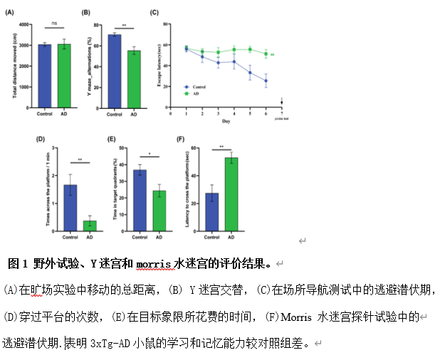
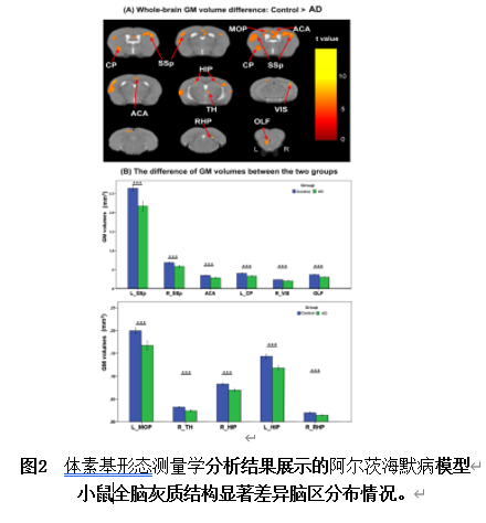
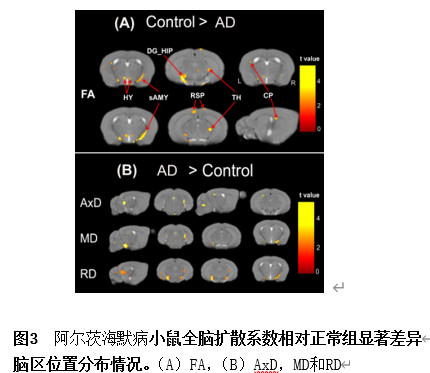
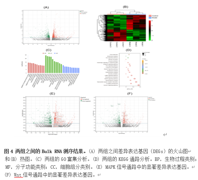
阿尔茨海默病三基因转基因小鼠模型(3xTg-AD)是一种广泛应用的小鼠模型,其表现出区域特异性的进行性β淀粉样蛋白和tau病理变化。除这些病理特征外,通过磁共振成像在3xTg-AD小鼠的晚期阶段(>12月龄)或早至2月龄时已观察到脑结构异常。然而,针对该模型在淀粉样β蛋白沉积广泛且tau病理学开始出现的阶段(约9月龄)进行MRI研究的文献极为有限。实验旨在通过评估9月龄3xTg-AD小鼠的脑形态测量学及微结构改变,深入理解这些特定病理特征背后的神经机制。采用基于体素的形态测量分析对T2加权图像进行分析,并采用基于体素的分析对弥散张量成像进行分析,以识别模型小鼠与对照组之间的差异。与对照组(与3xTg-AD小鼠同性別和品系)相比,3xTg-AD模型小鼠双侧海马、右颞叶、左脑干、左嗅球和皮层(双侧初级躯体感觉皮层、左次级运动皮层、右前穿质和视觉皮层)的灰质或体积显著减少。连接这些灰质区域的白质纤维束体积也显示出显著下降。全脑扩散张量成像分析检测到3xTg-AD小鼠双侧齿状回、右颞叶、左海马旁回、右小脑幕、左脑干和皮层(双侧后顶叶)的各向异性分数显著降低。此外,左侧海马和右小脑幕神经影像生物与行为表现具有显著相关性。海马区免疫荧光分析显示3xTg-AD小鼠淀粉样β蛋白斑块数量和p-Tau蛋白表达显著多于对照组,RNA测序分析发现阿尔茨海默病小鼠许多差异表达基因在突触结构、学习和记忆过程以及与神经髓鞘化过程相关的通路中显著富集。这些发现表明,在大量淀粉样β蛋白沉积和tau病理开始阶段,3xTg-AD小鼠出现了广泛的解剖学和微结构变化。这些结果为3xTg-AD小鼠模型在这一特定病理转变阶段内在的神经机制的理解提供新的见解,并为阿尔茨海默病的基础研究设计提供新的设计思路。


https://orcid.org/0000-0002-0914-6386 (Yongxin Li); https://orcid.org/0000-0002-9161-5435 (Zuocheng Qiu)

关键词: 3xTg-AD小鼠模型, 阿尔茨海默病, 脑体积, 脑-行为相关性, 扩散张量成像, 分数各向异性, 磁共振成像(MRI), RNA测序(RNA-Seq), 体素基形态测量学

Abstract: The triple transgenic mouse model of Alzheimer’s disease (3×Tg-AD) is a widely used model that exhibits region-dependent patterns of progressive amyloid-β and tau pathology. Although structural brain abnormalities on magnetic resonance imaging have been observed in 3×Tg-AD mice at later disease stages (> 12 months) and as early as 2 months, few studies have investigated changes in these mice during the stage with extensive amyloid-β deposition and onset of tau pathology (around 9 months). This study aimed to assess brain morphometry and microstructure alterations in 9 month-old 3×Tg-AD mice to better understand the neural mechanisms underlying these specific pathological features. Voxel-based analyses were employed on T2-weighted and diffusion tensor imaging to identify differences between 3×Tg-AD and control mice. Compared with controls, 3×Tg-AD mice exhibited lower gray matter volume in several regions including both hippocampal regions, the right thalamus, the left caudoputamen, and the cortex. Reduced white matter volume was observed in fiber tracts including the corpus callosum, internal capsule, stria terminalis, and olfactory tract. Whole-brain diffusion tensor imaging analysis revealed a significant decrease in fractional anisotropy and an increase in both radial and mean diffusivity within the left dentate gyrus of the hippocampal region and right striatum-like amygdala nuclei, with no significant difference in axial diffusivity. Correlation analyses demonstrated significant associations between behavioral performance measures, with both gray and white matter volumes within regions showing significant morphometric differences. Notably, behavioral performance also exhibited significant correlations with diffusion tensor imaging measures particularly within the left dentate gyrus of the hippocampal region and right striatum-like amygdala nuclei. Immunofluorescence analysis confirmed increased amyloid-β plaques and p-Tau protein expression in the hippocampal regions of 3×Tg-AD mice, which corroborated the magnetic resonance imaging findings. Transcriptome analysis in hippocampus tissue identified 1389 differentially expressed genes. Gene Ontology and Kyoto Encyclopedia of Genes and Genomes pathway analyses revealed that numerous differentially expressed genes were enriched in biological processes relevant to synapse structure, cognition, learning, and memory, with particular emphasis on Wnt and mitogen-activated protein kinase signaling pathways. Collectively, these findings suggest that intricate anatomical and microstructural alterations occur in 3×Tg-AD model mice at the onset of pathology around 9 months, potentially driven by gene expression alterations. Moreover, our results support the potential utility of brain volume and diffusion metrics as biomarkers for Alzheimer’s disease pathology, which could have significant implications for clinical diagnosis of Alzheimer’s disease patients.

Key words: 3×Tg-AD mouse model, Alzheimer’s disease, brain volume, brain–behavior correlation, diffusion tensor imaging, fractional anisotropy, magnetic resonance imaging, nerve regeneration, RNA sequencing, voxel-based morphometry