中国神经再生研究(英文版) ›› 2026, Vol. 21 ›› Issue (8): 3629-3640.doi: 10.4103/NRR.NRR-D-24-00968

• 原著:脑损伤修复保护与再生 • 上一篇    下一篇

肿瘤坏死因子α刺激基因6促进脑出血后血肿清除的作用及机制


  • 出版日期:2026-08-18 发布日期:2026-04-27

Tumor necrosis factor-α–stimulated gene 6 promotes hematoma clearance after intracerebral hemorrhage in a mouse model

Xia Liu1, 2, 3, #, Dabao Yao1, 3, #, Yunjie Li1, 3, Shiling Chen1, 3, Yingxin Tang1, 3, Jingyi Wang1, 3, Jingfei Yang3, 4, Jie Jing4, 5, Jiahui Wang1, 3,#br# Ge Zhang1, 3, Luwei Nie1, 3, Yangyang Feng1, 3, Gaigai Li1, 3, *, Zhouping Tang1, 3, *#br#   

  1. 1Department of Neurology, Tongji Hospital, Tongji Medical College, Huazhong University of Science and Technology, Wuhan, Hubei Province, China; 
    2Department of Neurology, Jingzhou Hospital, Yangtze University, Jingzhou, Hubei Province, China; 
    3Hubei Key Laboratory of Neural Injury and Functional Reconstruction, Huazhong University of Science and Technology, Wuhan, Hubei Province, China; 
    4Department of Nuclear Medicine, Tongji Hospital, Tongji Medical College, Huazhong University of Science and Technology, Wuhan, Hubei Province, China; 
    5Department of Neurology, Qilu Hospital, Shandong University, Jinan, Shandong Province, China
  • Online:2026-08-18 Published:2026-04-27
  • Contact: Zhouping Tang, MD, PhD, ddjtzp@163.com; Gaigai Li, MD, lgghuster@163.com.
  • Supported by:
    The study was supported by the National Natural Science Foundation of China, Nos. 92148206, 82071330 (both to ZT), 82201474 (to GL); and a grant from Tongji Hospital, No. 2022ZHFY01 (to ZT).

摘要:

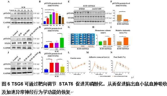
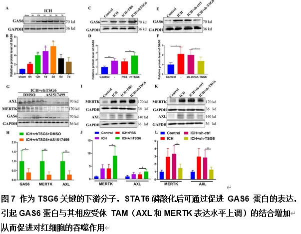
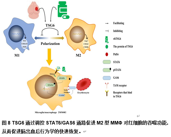
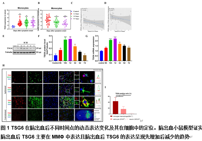
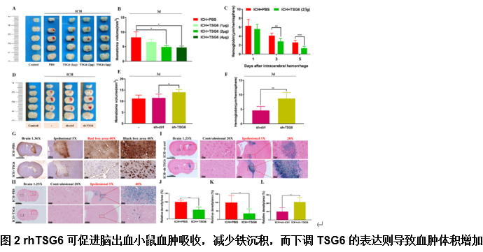
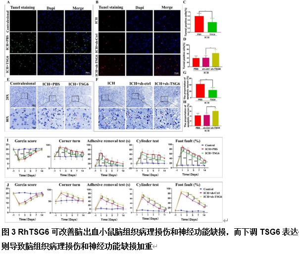
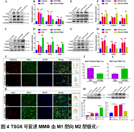
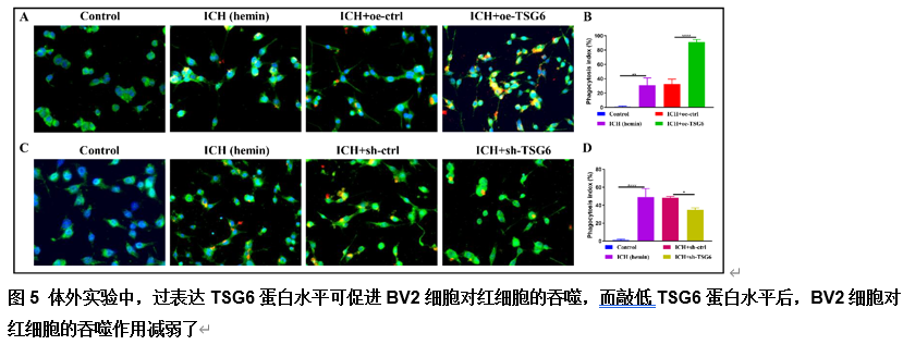
由于脑出血缺乏有效的治疗方法,其患者的预后较差。肿瘤坏死因子α刺激基因6(TSG6)是一种分泌型糖蛋白,可参与多种炎症性疾病的病理过程,并发挥抗炎作用。作者既往研究已发现,在体外脑出血模型中,脂肪来源的干细胞可通过上调TSG6蛋白来抑制炎症。但TSG6对体内血肿清除的直接作用机制仍未得到完全理解。实验探索了TSG6影响脑出血小鼠血肿吸收的潜在机制。实验首先分析了GEO数据库中脑出血患者的基因谱,并检测了脑出血小鼠脑组织中TSP6表达的变化,可见脑出血后TSG6表达呈一过性升高,且初始血肿体积与TSG6水平呈负相关。继而免疫荧光分析结果显示,TSG6主要在小胶质细胞/巨噬细胞中表达。随后发现TSG6可通过加速血肿清除、减少凋亡细胞和退化神经元、增加吞噬小胶质细胞/巨噬细胞的比例和减少铁沉积来促进脑出血小鼠功能的恢复。而后western blot和免疫荧光结果表明,TSG6可促进小胶质细胞/巨噬细胞的M2表型极化。体外吞噬实验进一步证明,TSG6可增强小胶质细胞吞噬红细胞的能力。最后发现实信号转导和转录激活因子6/生长停滞特异性蛋白6信号通路在TSG6介导的血肿吸收中的关键作用。综上,研究证实TSG6在促进脑出血小鼠模型中血肿吸收的关键作用,表明TSG6通过调节小胶质细胞/巨噬细胞细胞向M2型极化,激活信号转导和转录激活因子6/生长停滞特异性蛋白6信号通路,增加吞噬细胞表面相关吞噬受体表达增强其吞噬红细胞能力,从而加速血肿的清除并改善神经功能。


https://orcid.org/0000-0002-4153-8590 (Zhouping Tang); https://orcid.org/0000-0002-4083-8145 (Gaigai Li)

关键词: 脑出血, 血肿吸收, 肿瘤坏死因子α刺激基因6, 小胶质细胞/巨噬细胞, 极化, 信号转导子和转录激活子6, 生长停滞特异性蛋白6, 细胞凋亡, 炎症, 铁沉积

Abstract: The prognosis for patients who experience intracerebral hemorrhage is poor because of a lack of effective treatments. Tumor necrosis factor-α–stimulated gene 6 (TSG6) is a secreted glycoprotein that exerts anti-inflammatory effects in various inflammatory diseases. We previously showed that adipose-derived stem cells can inhibit inflammation by upregulating TSG6 secretion in an in vitro model of intracerebral hemorrhage. However, the direct effects of TSG6 on hematoma clearance in vivo remain largely unknown. The aim of this study was to determine how TSG6 affects hematoma absorption in mice subjected to intracerebral hemorrhage and to explore the potential underlying mechanisms. We first analyzed the gene profiles of patients with intracerebral hemorrhage from the GEO database and examined changes in TSG6 expression in the brain tissues of mice subjected to intracerebral hemorrhage. We found that TSG6 expression exhibited a transient increase following intracerebral hemorrhage, and that there was a negative correlation between the initial hematoma volume and TSG6 levels. Immunofluorescence analysis showed that TSG6 was primarily expressed in microglia and macrophages. Furthermore, we found that TSG6 promoted functional recovery in mice subjected to intracerebral hemorrhage by accelerating hematoma clearance, reducing the number of apoptotic cells and degenerated neurons, increasing the proportion of phagocytic microglia/macrophages, and decreasing iron deposition. Western blotting and immunofluorescence analysis indicated that TSG6 promoted M2 polarization of microglia/macrophages. In vitro phagocytosis experiments confirmed that TSG6 enhanced the ability of microglia to phagocytize red blood cells. Finally, we identified the signal transducer and activator of transcription 6/growth arrest–specific protein 6 signaling pathway as playing a critical role in TSG6-mediated hematoma absorption. In summary, our results demonstrate an essential role for TSG6 in promoting hematoma absorption in a mouse model of intracerebral hemorrhage. These findings suggest that TSG6 accelerates hematoma clearance and improves neurological function by promoting microglia/macrophage polarization to the M2 phenotype, activating the STAT6/GAS6 signaling pathway, and increasing phagocytic receptor expression on the surface of phagocytes, thereby enhancing their ability to phagocytize red blood cells.

Key words: apoptosis, growth arrest–specific protein 6, hematoma absorption, inflammation, intracerebral hemorrhage, iron deposition, microglia/macrophages, polarization, signal transducer and activator of transcription 6, tumor necrosis factor-α–stimulated gene 6