中国神经再生研究(英文版) ›› 2026, Vol. 21 ›› Issue (4): 1628-1640.doi: 10.4103/NRR.NRR-D-24-00461

• 原著:视神经损伤修复保护与再生 • 上一篇    下一篇

实验性青光眼Müller细胞内向整流钾通道Kir4.1和Kir4.1 Tyr9Asp突变过表达的神经保护作用


  • 出版日期:2026-04-15 发布日期:2025-07-28
  • 基金资助:
    本工作得到国家自然科学基金委、上海市科技重大项目、张江实验室等的资助。

Overexpression of the inwardly rectifying potassium channel Kir4.1 or Kir4.1 Tyr9 Asp in Müller cells exerts neuroprotective effects in an experimental glaucoma model

Fang Li1 , Zhen Li1 , Shuying Li1 , Hong Zhou1 , Yunhui Guo1 , Yongchen Wang2 , Bo Lei2, 3, Yanying Miao1, *, Zhongfeng Wang1, *   

  1. 1 State Key Laboratory of Medical Neurobiology and MOE Frontiers Center for Brain Science, Institute of Brain Science, Fudan University, Shanghai, China;  2 Institute of Neuroscience and Third Affiliated Hospital, Zhengzhou University, Zhengzhou, Henan Province, China;  3 Henan Provincial People’s Hospital, Henan Eye Institute, Henan Eye Hospital, People’s Hospital of Zhengzhou University, Zhengzhou University, Zhengzhou, Henan Province, China
  • Online:2026-04-15 Published:2025-07-28
  • Contact: Zhongfeng Wang, PhD, zfwang@fudan.edu.cn; Yanying Miao, PhD, yymiao@fudan.edu.cn.
  • Supported by:
    This study was supported by the National Natural Science Foundation of China, Nos. 32271043 (to ZW) and 82171047 (to YM); the both Science and Technology Major Project of Shanghai, No. 2018SHZDZX01 and ZJLab, and Shanghai Center for Brain Science and Brain-Inspired Technology (to ZW).

摘要:

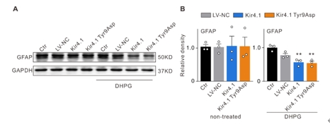
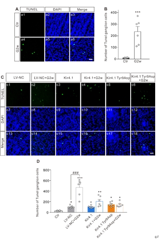
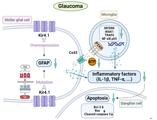
内向整流钾通道Kir4.1的表达和功能下调是诱导视网膜Müller细胞活化和神经胶质细胞相互作用的关键步骤,可参与青光眼视网膜神经节细胞的凋亡。因此调节Müller细胞中Kir4.1的表达可能是减轻青光眼视网膜神经节细胞损伤的潜在策略。此次实验中首先预测了Kir4.1蛋白中的7个磷酸化位点可发生突变,继而构建了含有不同突变位点的Kir4.1慢病毒表达载体,然后在mGluR I的激动剂DHPG处理以激活Müller胶质细胞的离体及在体模型中过表达Kir4.1和突变Kir4.1第9位酪氨酸,发现可抑制Müller胶质细胞的激活。接着在眼前房注射微磁珠建立的大鼠慢性高眼压动物模型中,发现Kir4.1过表达和Kir4.1 Tyr9Asp突变的Müller细胞中获得了类似的结果。过表达Kir4.1和Kir4.1 Tyr9Asp突变可抑制Müller胶质细胞激活,调控Bax/Bcl-2的平衡以及减少促炎因子白细胞介素1β和肿瘤坏死因子α的mRNA和蛋白质水平。进而在Müller胶质细胞和小胶质细胞共培养系统中研究了Müller胶质细胞Kir4.1过表达和Kir4.1 Tyr9Asp突变对促炎因子释放的调控作用,发现在共培养系统中,激活Müller细胞中ATP浓度和转运蛋白(小胶质细胞激活的标志物)水平升高,活化的Müler细胞诱导的小胶质细胞中炎症因子释放相关信号分子水平升高,上述变化可Kir4.1过表达和Kir4.1 Tyr9Asp突变所逆转。此外,Kir4.1过表达,而不是Kir4.1 Tyr9Asp突变,可减少活化的Müller细胞诱导的增殖和迁移小胶质细胞的数量。上述结果表明,在实验性青光眼视网膜中,Kir4.1 Tyr9可能是一个功能性调节位点。Kir4.1过表达和Kir4.1 Tyr9Asp突变减弱Müller细胞活化,减少ATP/P2X受体介导的神经胶质细胞相互作用,进而抑制小胶质细胞活化,降低促炎因子的合成和释放,从而减轻青光眼视网膜神经节细胞的凋亡。

https://orcid.org/0000-0002-2279-6885 (Zhongfeng Wang); https://orcid.org/0000-0003-3948-2943 (Yanying Miao)

关键词: Kir4.1过表达, Kir4.1 Tyr9Asp突变, Müller细胞, 小胶质细胞, 神经炎症, 神经保护, 视网膜神经节细胞, 慢性高眼压, 胶质细胞活化, 细胞凋亡

Abstract: Downregulation of the inwardly rectifying potassium channel Kir4.1 is a key step for inducing retinal Müller cell activation and interaction with other glial cells, which is involved in retinal ganglion cell apoptosis in glaucoma. Modulation of Kir4.1 expression in Müller cells may therefore be a potential strategy for attenuating retinal ganglion cell damage in glaucoma. In this study, we identified seven predicted phosphorylation sites in Kir4.1 and constructed lentiviral expression systems expressing Kir4.1 mutated at each site to prevent phosphorylation. Following this, we treated Müller glial cells in vitro and in vivo with the mGluR I agonist DHPG to induce Kir4.1 or Kir4.1 Tyr9 Asp overexpression. We found that both Kir4.1 and Kir4.1 Tyr9 Asp overexpression inhibited activation of Müller glial cells. Subsequently, we established a rat model of chronic ocular hypertension by injecting microbeads into the anterior chamber and overexpressed Kir4.1 or Kir4.1 Tyr9 Asp in the eye, and observed similar results in Müller cells in vivo as those seen in vitro. Both Kir4.1 and Kir4.1 Tyr9 Asp overexpression inhibited Müller cell activation, regulated the balance of Bax/Bcl-2, and reduced the mRNA and protein levels of pro-inflammatory factors, including interleukin-1β and tumor necrosis factor-α. Furthermore, we investigated the regulatory effects of Kir4.1 and Kir4.1 Tyr9 Asp overexpression on the release of pro-inflammatory factors in a co-culture system of Müller glial cells and microglia. In this co-culture system, we observed elevated adenosine triphosphate concentrations in activated Müller cells, increased levels of translocator protein (a marker of microglial activation), and elevated interleukin1β mRNA and protein levels in microglia induced by activated Müller cells. These changes could be reversed by Kir4.1 and Kir4.1 Tyr9 Asp overexpression in Müller cells. Kir4.1 overexpression, but not Kir4.1 Tyr9 Asp overexpression, reduced the number of proliferative and migratory microglia induced by activated Müller cells. Collectively, these results suggest that the tyrosine residue at position nine in Kir4.1 may serve as a functional modulation site in the retina in an experimental model of glaucoma. Kir4.1 and Kir4.1 Tyr9 Asp overexpression attenuated Müller cell activation, reduced ATP/P2X receptor–mediated interactions between glial cells, inhibited microglial activation, and decreased the synthesis and release of pro-inflammatory factors, consequently ameliorating retinal ganglion cell apoptosis in glaucoma.

Key words: apoptosis, chronic ocular hypertension, glial cell activation, Kir4.1 overexpression, Kir4.1 Tyr9 Asp mutation, microglia, Müller cells, neuroinflammation, neuroprotection, retinal ganglion cells