NRR:苏州大学丛启飞团队阐释突触修剪过程中小胶质细胞介导巡视和吞噬的分子机制
撰文:霍安冉,李奇,王佳丽,李梦琪,亓玉婉,尹俏,罗蔚锋,石际俊,丛启飞
小胶质细胞是大脑中重要的固有免疫细胞,在大脑的生理和病理状态中发挥重要作用。研究表明,小胶质细胞对周围的环境起着动态监视的功能。在大脑发育过程中,小胶质细胞可参与神经元环路连接的建立和成熟[1]。小胶质细胞还具有广泛的功能,包括调节神经发生[2]、诱导突触发生[3]、修剪不成熟突触[4]、促进神经元凋亡[5]和吞噬细胞碎片[6],也对中枢神经系统的吞噬、清除和免疫监视至关重要。
最近来自中国苏州大学神经科学研究所、苏州大学附属第二医院神经疾病研究中心丛启飞教授团队在《中国神经再生研究(英文版)》(Neural Regeneration Research)上发表了题为“Molecular mechanisms underlying microglial sensing and phagocytosis in synaptic pruning”的综述,该文系统地概述小胶质细胞介导的突触修剪的分子机制,同时探索了多种重要的免疫分子介导的信号通路在小胶质介导的突触修剪中的作用机制。
突触是神经元信息交流和传递的关键部位,对神经环路连接和功能发挥具有至关重要的作用。在这个过程中离不开突触形成、消除和重塑。首先,起始阶段需要神经轴突延伸至特定的部位;其次,轴突的末端开始进行了蛋白组装,形成一些膜蛋白或囊泡;再次,形成的突触连接经过突触可塑性的活性依赖方式得到增强;最后,再经活性依赖的方式对未成熟或功能弱的突触进行修剪[7](图1)。

图1 突触形成和消除(图源:Huo et al., Neural Regen Res, 2024)
小胶质细胞作为大脑中常驻免疫细胞,具有免疫调节和吞噬功能。小胶质细胞介导的突触修剪作用已在大脑发育的视网膜[8]、海马[4]和视觉皮质[9]中进行了广泛的研究。此外,近年来在其他脑区小胶质细胞介导突触修剪的作用也得到广泛关注。
突触修剪是由多种因素引起,包括了神经元内在因素和外在因素。近年来,胶质细胞作为外在因素在轴突修剪和突触消除中的作用逐渐被研究者所关注。丛启飞等主要关注小胶质细胞如何参与突触修剪过程,小胶质细胞介导的突触修剪包括了物理接触、吞噬和降解突触等过程,在这过程中,“找我”信号、“吃我”信号和“别吃我”信号发挥着重要作用(图2)。
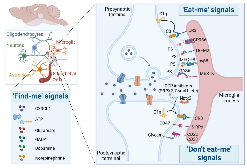
图2 “找我”、“吃我”和“别吃我”信号介导的小胶质细胞突触修剪作用(图源:Huo et al., Neural Regen Res, 2024)
以往研究表明,小胶质细胞通过配体与受体的相互作用,对微环境行使物理监视的作用[10-12]。研究表明,小胶质细胞表达多种神经递质受体,包括代谢型和离子型谷氨酸受体、γ-氨基丁酸受体、多巴胺受体、去甲肾上腺素受体、大麻素受体和乙酰胆碱受体、以及一些神经肽受体和神经调节剂如肾上腺素能受体和嘌呤受体[13]。
谷氨酸作为大脑中重要的神经递质,不仅可以从突触囊泡中释放,也可以从胶质细胞中释放。谷氨酸的过度释放可以刺激小胶质细胞的谷氨酸能受体,导致小胶质细胞激活产生活性氧,释放ATP和细胞因子,从而对树突棘减少和突触丢失产生对应的影响[14]。近期一项研究表明,代谢型谷氨酸能受体5的变构调节剂通过阻止小胶质吞噬补体C1q标记的突触来改善阿尔茨海默病小鼠的突触密度减少[15]。
γ-氨基丁酸是另一种重要的神经递质,由谷氨酸在中枢神经系统中通过谷氨酸脱羧酶合成,γ-氨基丁酸被包装到囊泡γ-氨基丁酸转运体中,释放到突触间隙中,与神经元γ-氨基丁酸A、γ-氨基丁酸C离子型受体、γ-氨基丁酸B代谢型受体和小胶质细胞的γ-氨基丁酸A/B受体结合[16]。以往的研究大多关注小胶质细胞介导的兴奋性突触修剪[8, 17]。最近的一项研究表明小胶质细胞可以通过γ-氨基丁酸信号选择性地修剪抑制性突触[12]。研究发现抑制性神经元释放神经递质γ-氨基丁酸,特异性地结合小胶质细胞表达的γ-氨基丁酸B受体,招募周围的小胶质细胞,触发一系列的复杂过程对未成熟的突触进行修剪。
多巴胺作为一种丰富的脑内神经递质,在大脑的不同脑区包括纹状体、前额叶皮质、下丘脑和黑质等起着重要的调节作用。神经元分泌的多巴胺与对应的受体相结合。近期的一项工作表明位于杏仁核的多巴胺能受体Drd2通过激活mTOR信号通路调控突触修剪[18]。研究发现,伏隔核小胶质细胞表达的多巴胺能受体Drd1参与调控补体C3-C3R介导的小胶质细胞吞噬,从而在社会交互行为中发挥重要作用。
其他一些神经递质,比如去甲肾上腺素以及乙酰胆碱,在参与小胶质细胞介导的突触修剪过程中还有待进一步研究。
此外,神经元还分泌其他一些重要的“找我”信号分子,包括趋化因子和ATP。这些信号分子可以结合小胶质细胞表面受体,激活小胶质细胞从而募集小胶质细胞迁移至突触周围,调控突触可塑性[19-22]。
小胶质细胞介导的突触修剪主要包括三个过程即识别、吞噬和消化[23]。通过一些“吃我”信号比如磷脂酰丝氨酸、钙网蛋白、Gas6和补体蛋白结合小胶质细胞表面相应的吞噬受体[23],特异性启动受体介导的信号传导,诱导细胞骨架蛋白重塑,吞噬突触,最终通过吞噬体与溶酶体融合导致突触被消化。
目前研究广泛的“吃我”信号是磷脂酰丝氨酸,其暴露在死亡神经元和活性弱的突触上,与调理素及其相关分子结合后作用于小胶质细胞αvβ5整合素受体[24, 25],诱导凋亡细胞的吞噬[24, 26]。此外,研究发现黏附G蛋白偶联受体56可以通过特定结构域结合磷脂酰丝氨酸。小胶质细胞特异性缺失G蛋白偶联受体56,导致小胶质细胞吞噬磷脂酰丝氨酸标记的突触减少,显著增加囊泡谷氨酸转运体2兴奋性突触密度[27]。
补体系统是重要的天然免疫组成部分,补体蛋白C1q、C3和C4介导的小胶质细胞吞噬在大脑神经突触修剪中的作用已得到广泛研究[8, 28]。但目前仍存在一些尚未解决的问题,比如补体蛋白是否是广泛的突触重塑调控分子,大脑如何调控补体介导小胶质细胞突触修剪作用。
近年来研究发现,大脑中存在着一些“别吃我”信号负责调控小胶质细胞介导的突触修剪作用[29]。SRPX2是一种新发现主要由神经元分泌的经典补体通路调节蛋白, SRPX2的缺失会破坏补体蛋白介导的小胶质突触修剪的平衡,导致发育中的外侧膝状体区和体感皮质区中囊泡谷氨酸转运体2兴奋性突触密度的减少[30]。近期另一项研究发现了一种新型补体调节蛋白Nptx2,其可抑制经典补体通路活性,并调控小鼠大脑兴奋性突触密度[31]。此外,还发现一些含有sushi结构域蛋白在突触形成和突触可塑性调控中发挥着作用[32, 33],但这些蛋白是否通过调节补体介导的小胶质细胞吞噬在突触修剪中发挥重要作用尚不清楚。
CD47-SIRPα信号是另一种重要的“别吃我”信号,在大脑发育和神经退行性病变中保护突触免受小胶质细胞过度修剪突触。研究发现CD47的缺失导致突触过度修剪[34],并且阿尔茨海默病小鼠模型中的小胶质细胞特异性的SIRPα缺失加速了突触丢失[35]。
此外,一些糖基化修饰蛋白,特别是唾液酸化的蛋白,在小胶质细胞介导的突触稳态调节中起着至关重要的作用。神经元唾液酸化蛋白通过与小胶质细胞表面的唾液酸结合免疫球蛋白样凝集素的相互作用抑制小胶质细胞吞噬[36, 37],神经元膜蛋白上的唾液酸糖基参与补体蛋白C1q与突触的结合,保护神经突触免受补体CR3介导的小胶质细胞吞噬[38]。
上述“找我”、“吃我”和“别吃我”信号与免疫系统密不可分[39]。近年来研究发现,一些免疫信号分子也参与了小胶质细胞调控突触修剪的过程(图3)。

图3 免疫相关信号分子介导的小胶质细胞突触修剪作用(图源:Huo et al., Neural Regen Res, 2024)
CX3CL1作为一种神经元分泌的趋化因子,作用于小胶质细胞表面CX3CR1受体,特异性敲除CX3CR1基因导致小胶质细胞数量短暂减少,延缓突触修剪作用[4]。TREM2是另一种与吞噬功能相关的小胶质细胞特异性免疫受体,是阿尔茨海默病的一个危险因素[40]。TREM2缺失导致突触密度增加,同时伴有兴奋性突触传递增强[41]。
另外,星形胶质细胞分泌细胞因子白细胞介素33能够将小胶质细胞招募到冗余突触附近。缺乏白细胞介素33的小鼠在发育过程中表现出兴奋性突触增加,小胶质细胞吞噬功能异常[42]。报道发现白细胞介素33/ST2信号通路介导小胶质细胞-神经突触相互作用在调控海马突触可塑性和突触重塑中起着关键作用[43]。
JAK2-STAT1是经典的免疫调节信号通路,JAK2的激活促进STAT1磷酸化。JAK2-STAT1信号通路的失活对突触修剪具有保护作用[44],但是JAK2-STAT1信号通路是否调控小胶质细胞吞噬功能仍需进一步研究。
首先,此次综述聚焦在3种不同信号分子介导的小胶质细胞对微环境的响应及应答,而这种响应与应答并不仅限于神经元与小胶质细胞的相互作用,越来越多的研究表明星形胶质细胞也参与了突触修剪过程。此外,文章没有详细阐述不同状态的小胶质细胞对于突触修剪的差异。
小胶质细胞是大脑中的常驻免疫细胞,在大脑发育和神经回路连接中发挥重要作用。小胶质细胞及其相关免疫分子是突触形成和消除所必需的,也是神经网络发育中突触正确连接的关键。小胶质细胞对来自神经元的“找我”信号和来自周围微环境的“吃我”或“别吃我”信号的响应,监测神经元活动,对兴奋性突触或抑制性突触进行特异性修剪。通过揭示神经元-小胶质细胞相互作用在突触修剪中的分子机制,无疑为小胶质细胞在发育到疾病的突触修剪功能提供新的思路,同时为药物靶点发现和开发针对突触功能障碍的神经系统疾病治疗提供重要线索。
原文链接:https://doi.org/10.4103/1673-5374.385854
参考文献
[1] Salter MW, Beggs S. Sublime microglia: expanding roles for the guardians of the CNS. Cell. 2014;158(1):15-24.
[2] Sato K. Effects of Microglia on Neurogenesis. Glia. 2015;63(8):1394-1405.
[3] Miyamoto A, Wake H, Ishikawa AW, et al. Microglia contact induces synapse formation in developing somatosensory cortex. Nat Commun. 2016;7:12540.
[4] Paolicelli RC, Bolasco G, Pagani F, et al. Synaptic pruning by microglia is necessary for normal brain development. Science. 2011;333(6048):1456-1458.
[5] Anderson SR, Roberts JM, Ghena N, et al. Neuronal apoptosis drives remodeling states of microglia and shifts in survival pathway dependence. Elife. 2022;11:e76564.
[6] Bejarano-Escobar R, Sánchez-Calderón H, Otero-Arenas J, et al. Müller glia and phagocytosis of cell debris in retinal tissue. J Anat. 2017;231(4):471-483.
[7] Fourgeaud L, Boulanger LM. Synapse remodeling, compliments of the complement system. Cell. 2007;131(6):1034-1036.
[8] Schafer DP, Lehrman EK, Kautzman AG, et al. Microglia sculpt postnatal neural circuits in an activity and complement-dependent manner. Neuron. 2012;74(4):691-705.
[9] Tremblay M, Lowery RL, Majewska AK. Microglial interactions with synapses are modulated by visual experience. PLoS Biol. 2010;8(11):e1000527.
[10] Davalos D, Grutzendler J, Yang G, et al. ATP mediates rapid microglial response to local brain injury in vivo. Nat Neurosci. 2005;8(6):752-758.
[11] Haynes SE, Hollopeter G, Yang G, et al. The P2Y12 receptor regulates microglial activation by extracellular nucleotides. Nat Neurosci. 2006;9(12):1512-1519.
[12] Favuzzi E, Huang S, Saldi GA, et al. GABA-receptive microglia selectively sculpt developing inhibitory circuits. Cell. 2021;184:4048-4063.e32(15).
[13] Liu H, Leak RK, Hu X. Neurotransmitter receptors on microglia. Stroke Vasc Neurol. 2016;1(2):52-58.
[14] Haroon E, Miller AH, Sanacora G. Inflammation, Glutamate, and Glia: A Trio of Trouble in Mood Disorders. Neuropsychopharmacology. 2017;42(1):193-215.
[15] Spurrier J, Nicholson L, Fang XT, et al. Reversal of synapse loss in Alzheimer mouse models by targeting mGluR5 to prevent synaptic tagging by C1Q. Sci Transl Med. 2022;14(647):eabi8593.
[16] Luján R, Shigemoto R, López-Bendito G. Glutamate and GABA receptor signalling in the developing brain. Neuroscience. 2005;130(3):567-580.
[17] Gunner G, Cheadle L, Johnson KM, et al. Sensory lesioning induces microglial synapse elimination via ADAM10 and fractalkine signaling. Nat Neurosci. 2019;22(7):1075-1088.
[18] Zhang YQ, Lin WP, Huang LP, et al. Dopamine D2 receptor regulates cortical synaptic pruning in rodents. Nat Commun. 2021;12(1):6444.
[19] Flagmeyer I, Haas HL, Stevens DR. Adenosine A1 receptor-mediated depression of corticostriatal and thalamostriatal glutamatergic synaptic potentials in vitro. Brain Res. 1997;778(1):178-185.
[20] Yabuuchi K, Kuroiwa M, Shuto T, et al. Role of adenosine A1 receptors in the modulation of dopamine D1 and adenosine A2A receptor signaling in the neostriatum. Neuroscience. 2006;141(1):19-25.
[21] Trusel M, Cavaccini A, Gritti M, et al. Coordinated Regulation of Synaptic Plasticity at Striatopallidal and Striatonigral Neurons Orchestrates Motor Control. Cell Rep. 2015;13(7):1353-1365.
[22] Corkrum M, Covelo A, Lines J, et al. Dopamine-Evoked Synaptic Regulation in the Nucleus Accumbens Requires Astrocyte Activity. Neuron. 2020;105:1036-1047.e5(6).
[23] Vilalta A, Brown GC. Neurophagy, the phagocytosis of live neurons and synapses by glia, contributes to brain development and disease. FEBS J. 2018;285(19):3566-3575.
[24] Albert ML, Kim JI, Birge RB. alphavbeta5 integrin recruits the CrkII-Dock180-rac1 complex for phagocytosis of apoptotic cells. Nat Cell Biol. 2000;2(12):899-905.
[25] Lian H, Yang L, Cole A, et al. NFκB-activated astroglial release of complement C3 compromises neuronal morphology and function associated with Alzheimer's disease. Neuron. 2015;85(1):101-115.
[26] Akakura S, Singh S, Spataro M, et al. The opsonin MFG-E8 is a ligand for the alphavbeta5 integrin and triggers DOCK180-dependent Rac1 activation for the phagocytosis of apoptotic cells. Exp Cell Res. 2004;292(2):403-416.
[27] Li T, Chiou B, Gilman CK, et al. A splicing isoform of GPR56 mediates microglial synaptic refinement via phosphatidylserine binding. EMBO J. 2020;39(16):e104136.
[28] Stevens B, Allen NJ, Vazquez LE, et al. The classical complement cascade mediates CNS synapse elimination. Cell. 2007;131(6):1164-1178.
[29] Wang C, Wang L, Gu Y. Microglia, synaptic dynamics and forgetting. Brain Res Bull. 2021;174:173-183.
[30] Cong Q, Soteros BM, Wollet M, et al. The endogenous neuronal complement inhibitor SRPX2 protects against complement-mediated synapse elimination during development. Nat Neurosci. 2020;23(9):1067-1078.
[31] Zhou J, Wade SD, Graykowski D, et al. The neuronal pentraxin Nptx2 regulates complement activity and restrains microglia-mediated synapse loss in neurodegeneration. Sci Transl Med. 2023;15(689):eadf0141.
[32] Nadjar Y, Triller A, Bessereau JL, et al. The Susd2 protein regulates neurite growth and excitatory synaptic density in hippocampal cultures. Mol Cell Neurosci. 2015;65:82-91.
[33] González-Calvo I, Iyer K, Carquin M, et al. Sushi domain-containing protein 4 controls synaptic plasticity and motor learning. Elife. 2021;10:e65712.
[34] Lehrman EK, Wilton DK, Litvina EY, et al. CD47 Protects Synapses from Excess Microglia-Mediated Pruning during Development. Neuron. 2018;100:120-134.e6(1).
[35] Ding X, Wang J, Huang M, et al. Loss of microglial SIRPα promotes synaptic pruning in preclinical models of neurodegeneration. Nat Commun. 2021;12(1):2030.
[36] Wang Y, Neumann H. Alleviation of neurotoxicity by microglial human Siglec-11. J Neurosci. 2010;30(9):3482-3488.
[37] Claude J, Linnartz-Gerlach B, Kudin AP, et al. Microglial CD33-related Siglec-E inhibits neurotoxicity by preventing the phagocytosis-associated oxidative burst. J Neurosci. 2013;33(46):18270-18276.
[38] Linnartz B, Kopatz J, Tenner AJ, et al. Sialic acid on the neuronal glycocalyx prevents complement C1 binding and complement receptor-3-mediated removal by microglia. J Neurosci. 2012;32(3):946-952.
[39] Wu Y, Dissing-Olesen L, Macvicar BA, et al. Microglia: Dynamic Mediators of Synapse Development and Plasticity. Trends Immunol. 2015;36(10):605-613.
[40] Zhou Y, Song WM, Andhey PS, et al. Human and mouse single-nucleus transcriptomics reveal TREM2-dependent and TREM2-independent cellular responses in Alzheimer's disease. Nat Med. 2020;26(1):131-142.
[41] Filipello F, Morini R, Corradini I, et al. The Microglial Innate Immune Receptor TREM2 Is Required for Synapse Elimination and Normal Brain Connectivity. Immunity. 2018;48:979-991.e8(5).
[42] Vainchtein ID, Chin G, Cho FS, et al. Astrocyte-derived interleukin-33 promotes microglial synapse engulfment and neural circuit development. Science. 2018;359(6381):1269-1273.
[43] Wang Y, Fu AKY, Ip NY. IL-33/ST2 Signaling Regulates Synaptic Plasticity and Homeostasis in the Adult Hippocampal Circuitry. DNA Cell Biol. 2021;40(9):1125-1130.
[44] Yasuda M, Nagappan-Chettiar S, Johnson-Venkatesh EM, et al. An activity-dependent determinant of synapse elimination in the mammalian brain. Neuron. 2021;109:1333-1349.e6(8).
 #br#
#br#
第一作者:霍安冉,苏州大学神经科学研究所2021级在读硕士。
 #br#
#br#
通讯作者:丛启飞,现任苏州大学教授,博士研究生导师。主要研究神经元与小胶质细胞互作的分子机制,尤其是神经突触形成、修剪和再生的免疫调控机制,聚焦于突触功能异常导致的精神类疾病的病理机制和干预策略研究。在国际学术期刊Nature Neuroscience、Glia、Acta Pharmacologica Sinica等期刊发表14篇学术论文。担任Brain-X期刊青年编委,入选2023年度苏州市科协青年科技人才托举工程。
 #br#
#br#
丛启飞团队成员合影