NRR:重庆医科大学肖农团队分析脊髓损伤后小胶质细胞与巨噬细胞不同的动态演化过程
撰文:周虎耀,王霞,肖农,黎国兴
脊髓损伤会导致永久的运动、感觉与自主神经障碍,而这种损伤一般可被分为2个阶段:以细胞缺血、水肿、氧化应激为主的原发性损伤以及以原位细胞激活、免疫细胞浸润等为主的继发性损伤[1, 2]。常驻的小胶质细胞与浸润的巨噬细胞都在神经炎症中扮演着重要的角色。由于2种细胞结构与功能的相似性,许多研究者将其归为一类,并分为促炎的M1亚型与抗炎的M2亚型[3, 4]。然而,越来越多的研究表明,小胶质细胞与巨噬细胞仍存在着一定差异[5],前者主要参与中枢神经系统稳态维持、神经元兴奋性调节以及髓鞘再生与突触剪切,而后者主要参与急性期炎症反应与组织修复。在阿尔茨海默病中,2种细胞对疾病进程有着不同的影响[6]。目前,小胶质细胞与巨噬细胞对脊髓损伤后的独立作用尚不明确,对其探索可以进一步地完善脊髓损伤后的病理机制,并发现潜在的治疗靶点。
中国重庆医科大学肖农团队在《中国神经再生研究(英文)》(Neural Regeneration Research)上发表了题为“Dynamic development of microglia and macrophages after spinal cord injury”的研究,其通过生信分析结合部分体内实验,探索了脊髓损伤后小胶质细胞与巨噬细胞不同的演化进程。小胶质细胞在损伤后立即激活为促炎的表型,并随着疾病进展逐渐转化为抗炎的稳定态。而巨噬细胞在损伤后同时具有促炎和神经保护作用,并随着疾病进展均逐渐减少,其中促炎作用可能与整合素β2(Itgb2)有关,而神经保护作用可能与制瘤素 M(Osm)通路有关。该研究可对脊髓损伤后的炎症细胞干预提供新的思路。
肖农等通过Gene Expression Omnibus database公共数据库获得脊髓损伤后不同时间的单细胞数据后,成功进行了细胞的降维与分群。实验首先关注了小胶质细胞群体,雷达图以及基因动力学分析均提示了其在脊髓损伤后从促炎态向稳定态的转变(图1)。为进一步探索炎症进程,肖农等进行细胞间通讯分析与体内实验,发现Osm通路的显著上调,使用网络图提示了该通路在巨噬细胞中的主要表达;结合配体-受体配对分析以及空间转录组,研究者发现其可能作用于神经元上的Rack1受体(图2),而既往研究提示该受体的激活具有神经保护作用[7]。因此,单独关注巨噬细胞群体,提示了促炎基因与修复基因在损伤后第1天的同步高表达,而基因动力学和空间转录组提示了Itgb2对巨噬细胞命运调控的重要性,Itgb2在既往研究中更多的被认为与炎症相关[8],故进一步使用体内实验明确了其在脊髓损伤后其随时间的下降(图3)。这些结果提示小胶质细胞与巨噬细胞在脊髓损伤可能扮演角色的不同。
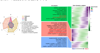
图1脊髓损伤后小胶质细胞促炎态向稳态的演化(图源:Wang et al.,Neural Regen Res, 2025)
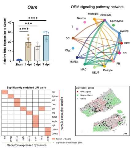
图2脊髓损伤后巨噬细胞与Osm通路分析(图源:Wang et al.,Neural Regen Res, 2025)
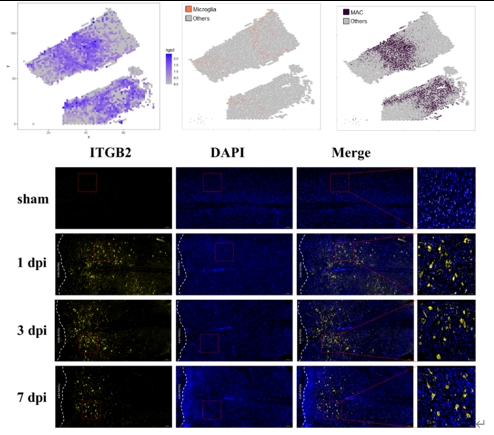
图3脊髓损伤后Itgb2表达情况(图源:Zhou et al.,Neural Regen Res, 2025)
总之,肖农等提出脊髓损伤后小胶质细胞存在着从促炎向抗炎的转化,而巨噬细胞在早期就同时具有促炎和神经保护的功能,并均随着时间逐渐下降,其中神经保护作用可能与OSM通路有关,而促炎作用可能与Itgb2有关。该研究对脊髓损伤后炎症反应的调控方式提供新的思路。然而,要明确这个假设,还需要更多的体内外实验进行佐证;其次,该研究运用的数据集为单细胞测序而非单细胞核测序,因此未分析星形胶质细胞相关的数据,对神经元的单细胞分析并非最佳;最后,该研究仅关注了7d内的炎症反应,长期的炎症演化也还值得进一步探索。
原文链接:https://doi.org/10.4103/NRR.NRR-D-24-00063
参考文献
[1] Hellenbrand DJ, Quinn CM, Piper ZJ, et al. Inflammation after spinal cord injury: a review of the critical timeline of signaling cues and cellular infiltration. J Neuroinflammation. 2021;18(1):284.
[2] Silva NA, Sousa N, Reis RL, et al. From basics to clinical: a comprehensive review on spinal cord injury. Prog Neurobiol. 2014;114:25-57.
[3] Wu H, Zheng J, Xu S, et al. Mer regulates microglial/macrophage M1/M2 polarization and alleviates neuroinflammation following traumatic brain injury. J Neuroinflammation. 2021;18(1):2.
[4] Kumar A, Barrett JP, Alvarez-Croda DM, et al. NOX2 drives M1-like microglial/macrophage activation and neurodegeneration following experimental traumatic brain injury. Brain Behav Immun. 2016;58:291-309.
[5] Sevenich L. Brain-resident microglia and blood-borne macrophages orchestrate central nervous system inflammation in neurodegenerative disorders and brain cancer. Front Immunol. 2018;9:697.
[6] Prinz M, Erny D, Hagemeyer N. Ontogeny and homeostasis of CNS myeloid cells. Nat Immunol. 2017;18(4):385-392.
[7] Ni H, Rui Q, Xu Y, et al. RACK1 upregulation induces neuroprotection by activating the IRE1-XBP1 signaling pathway following traumatic brain injury in rats. Exp Neurol. 2018;304:102-113.
[8] Bhowmick S, Malat A, Caruso D, et al. Intercellular adhesion molecule-1-induced posttraumatic brain injury neuropathology in the prefrontal cortex and hippocampus leads to sensorimotor function deficits and psychological stress. eNeuro. 2021;8 (4):ENEURO.0242-21.2021.