中国神经再生研究(英文版) ›› 2026, Vol. 21 ›› Issue (6): 2506-2513.doi: 10.4103/NRR.NRR-D-24-00974

• 原著:脊髓损伤修复保护与再生 • 上一篇    下一篇

食蟹猴脊髓损伤后背根神经节单细胞测序图谱:揭示中枢神经系统的细胞免疫微环境


  • 出版日期:2026-06-15 发布日期:2026-04-18

Single-cell RNA sequencing of the post–spinal cord injury dorsal root ganglia in cynomolgus monkeys: Elucidation of the cellular immune microenvironment of the central nervous system

Yiming Ren1, 2, #, Bo Li3, #, Bo Yang1, #, Baoyou Fan1, #, Shenghui Huang1, Guidong Shi4, Liang Liu3, *, Zhijian Wei4, *, Shiqing Feng1, 4, *   

  1. 1Department of Orthopedics, International Science and Technology Cooperation Base of Spinal Cord Injury, Tianjin Key Laboratory of Spine and Spinal Cord Injury, Tianjin Medical University General Hospital, Tianjin, China; 
    2Department of Joint and Sport Medicine, Tianjin Union Medical Center, The First Affiliated Hospital of Nankai University, Tianjin, China; 
    3Department of Orthopedics, Beijing Luhe Hospital, Capital Medical University, Beijing, China; 
    4Department of Othopedics, Qilu Hospital of Shandong University, Jinan, Shandong Province, China
  • Online:2026-06-15 Published:2026-04-18
  • Contact: Shiqing Feng, PhD, sqfeng@tmu.edu.cn; Zhijian Wei, PhD, weizhijian2002@126.com; Liang Liu, PhD, liuliang@ccmu.edu.cn.
  • Supported by:
    This study was supported by the Tianjin Key Medical Discipline (Specialty) Construct Project, No. TJYXZDXK-027A (to SF); the National Key Research and Development Project of Stem Cell and Transformation Research, No. 2019YFA0112100 (to SF); Tianjin Natural Science Foundation’s Youth Project for Diverse Investments, No. 21JCQNJC01300 (to BF); the National Natural Science Foundation of China (Youth Program), No. 82102563 (to BF); Tianjin Major Science and Technology Special Projects and Engineering Projects, No. 21ZXJBSY00080 (to YR).

摘要:

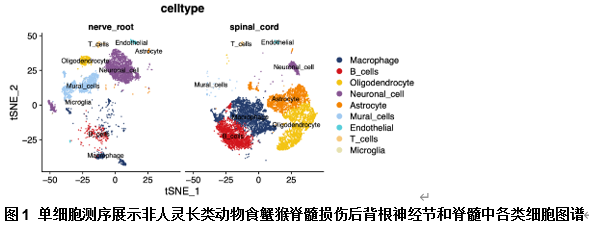
很少有研究关注脊髓损伤后背根神经节免疫细胞微环境的变化,以及这些变化是否有助于轴突再生。因此,此次实验采用单细胞RNA测序构建食蟹猴胸中段脊髓挫伤模型中背根神经节和脊髓中不同细胞类型的数据集。细胞通讯分析表明,特定的信号事件发生在多种背根神经节细胞类型中,以响应脊髓损伤。降维聚类的单细胞分析鉴定出9种细胞类型的不同分子特征。这些包括脊髓损伤后背根神经节细胞与脊髓细胞中的巨噬细胞亚型和基因表达谱。巨噬细胞亚群根据差异表达基因分为11个簇,标记为MC0-MC10,包含前10种基因,分别为ABCA6,RBMS3,EBF1,LAMA4,ANTXR2,LAMA2,SOX5,FOXP2,GHR和APOD。其中MC0,MC1和MC2构成了主要的巨噬细胞群,而MC4,MC6和MC9在脊髓中几乎不存在,但背根神经节显著增加。同时,作为多功能亚型,它们具有丰富的调节轴突再生的能力。通过细胞轨迹和伪时间分析,阐明了脊髓损伤后背根神经节中巨噬细胞的发育进程。同时,基因EBF1(MC6和MC9标记物)、RBMS3(MC6和MC9标记物)和ABCA6(MC6标记物)在巨噬细胞的关键通路中存在高表达。配体-受体对分析确定了巨噬细胞对小胶质细胞的影响,发现巨噬细胞主要通过SPP1-CD44,LAMC1-CD44和FN1-CD44相互作用,可能促进免疫微环境中的特异性细胞通讯。综上,实验首次全面分析了食蟹猴脊髓损伤后背根神经节的细胞异质性图谱,其涵盖了背根神经节区域内几乎所有细胞类型。基于该数据集评估了脊髓损伤后背根神经节区域内巨噬细胞的多种亚型,并检查了促进免疫反应相关巨噬细胞和小胶质细胞在背根神经节部位相互作用的信号通路。该研究为理解免疫微环境如何影响脊髓损伤后背根神经节中神经元的再生能力提供了理论基础,并提供了新的见解,其有助于解码脊髓损伤病理生理学的复杂过程。


https://orcid.org/0000-0001-9437-7674 (Shiqing Feng)

关键词: 脊髓损伤, 背根神经节, 单细胞序列, 巨噬细胞, 细胞微环境, 差异表达基因, 细胞通信, 小胶质细胞, 免疫细胞, 神经元

Abstract: Few studies have investigated alterations in the immune cell microenvironment of the dorsal root ganglia following spinal cord injury and whether these modifications facilitate axonal regeneration. In this study, we used a single-cell RNA sequencing dataset to create a comprehensive profile of the diverse cell types in the dorsal root ganglia and spinal cord of a mid-thoracic contusion injury model in cynomolgus monkeys. Cell communication analysis indicated that specific signaling events among various dorsal root ganglia cell types occur in response to spinal cord injury. Single-cell analysis using dimensionality reduction clustering identified distinct molecular signatures for nine cell types, including macrophage subpopulations, and differential gene expression profiles between dorsal root ganglia cells and spinal cord cells following spinal cord injury. The macrophage subpopulations were categorized into 11 clusters (MC0–MC10) based on differentially expressed genes, with the top 10 genes being ABCA6, RBMS3, EBF1, LAMA4, ANTXR2, LAMA2, SOX5, FOXP2, GHR, and APOD. MC0, MC1, and MC2 constituted the predominant macrophage populations. MC4, MC6, and MC9 were nearly absent in the spinal cord, but exhibited significant increases in the dorsal root ganglia post–spinal cord injury. Notably, these subpopulations possess a strong capacity for regulating axonal regeneration. The developmental progression of dorsal root ganglia macrophages after spinal cord injury was elucidated using cell trajectory and pseudo-time analyses. Genes such as EBF1 (MC6 and MC9 marker), RBMS3 (MC6 and MC9 marker), and ABCA6 (MC6 marker) showed high expression levels in the critical pathways of macrophage function. Through ligand–receptor pair analysis, we determined that the effects of macrophages on microglia are predominantly mediated through interaction pairs (e.g., SPP1-CD44, LAMC1-CD44, and FN1-CD44), potentially facilitating specific cellular communications within the immune microenvironment. The single-cell RNA sequencing dataset used in this study represents the first comprehensive transcriptional analysis of the dorsal root ganglia after spinal cord injury in cynomolgus monkeys, encompassing nearly all cell types within the dorsal root ganglia region. Using this dataset, we evaluated diverse subtypes of macrophages in the post- spinal cord injury dorsal root ganglia area and examined the signaling pathways that facilitate interactions among immune response-related macrophages in the dorsal root ganglia. Findings from this study provide a theoretical basis for understanding how the immune microenvironment influences the regenerative capacity of dorsal root ganglia neurons after spinal cord injury and offer novel insights into the complex processes underlying the pathobiology of spinal cord injury. 

Key words: cellular communication, cellular microenvironment, differentially expressed genes, dorsal root ganglia, immune cells, macrophage, microglia, neurons, single-cell sequence, spinal cord injury