NRR:中南大学王志远、刘军和卢伟团队揭示视神经脊髓炎谱系疾病无明显脑病灶患者大脑皮质改变与临床表现的关系
撰文:黄楚欣、李彦宇、陈雁菁、廖旋、张会婷、王志远、刘军、卢伟
视神经脊髓炎谱系疾病是一组中枢神经系统自身免疫性炎症性脱髓鞘疾病,全球发病率约为0.5-4/10万人[1]。视神经脊髓炎谱系疾病通常累及脊髓、视神经以及水通道蛋白4表达水平较高的脑区(如第三、四脑室、下丘脑和极后区)。尽管视神经脊髓炎谱系疾病患者可能会发生脑部病变,但仍存在部分患者在常规脑部磁共振成像下表现为正常或非特异性点片状白质改变的现象[2]。因此常规磁共振成像可能无法检测和量化某些隐匿性改变,已有多项研究使用不同的神经成像方式进行脑微观结构改变的探索。
既往研究表明,多发性硬化患者的皮质变薄与脑白质病变大小相关,这表明皮质萎缩可能是轴突损伤影响高度连接脑区的结果[3, 4]。另一项研究表明,伴有脑损伤的视神经脊髓炎谱系疾病患者表现出比无明显病灶的视神经脊髓炎谱系疾病患者及健康对照者更严重的丘脑萎缩[5]。因此推测,存在脑部病灶有可能会加重视神经脊髓炎谱系疾病的皮质萎缩,但无明显脑病灶的视神经脊髓炎谱系疾病患者是否出现皮质萎缩、其皮质改变特征以及其临床相关性目前尚不完全明确。
来自中国中南大学王志远、刘军和卢伟团队最近在《中国神经再生研究(英文版)》(Neural Regeneration Research)上发表了题为“Correlation between cerebral cortex changes and clinical features in patients with neuromyelitis optica spectrum disorder with normal-appearing brain tissue: a case-control study”的研究。他们发现,无明显脑部病灶的视神经脊髓炎谱系疾病患者比健康对照的双侧额中回喙部及额上回皮质更薄,且存在执行功能、工作记忆等方面的认知功能减退。进一步研究发现,视神经脊髓炎谱系疾病患者双侧额中回喙部的皮质厚度与其临床残疾和认知功能有相关性。这启示,对视神经脊髓炎谱系疾病患者的脑影像进行基于表面形态学分析的脑皮质微结构研究,可能为患者的临床表现及其相关机制提供相应的影像学解释,并为今后的个体化治疗提供一定的理论基础。
王志远等的研究纳入43例无明显脑部病灶的视神经脊髓炎谱系疾病患者和45名健康对照者。在所有视神经脊髓炎谱系疾病患者中,7例患者因视力障碍或上肢乏力而未能完成认知测试,其余患者的数字符号转换测试、连线测试A和连线测试B表现明显差于健康对照者(P < 0.001)。与健康对照组相比,视神经脊髓炎谱系疾病组双侧额中回喙部和左额上回的皮质厚度显著降低(P < 0.001, 团块水平FDR校正,图1)。而2组脑回指数和脑沟深度无显著差异。作者进一步以视神经炎临床发作作为脑病灶表现,比较了既往有视神经炎临床发作(n=19)与没有视神经炎临床发作的视神经脊髓炎谱系疾病患者(n=24)是否存在皮质厚度的差异。结果发现,与没有视神经炎临床发作的视神经脊髓炎谱系疾病患者相比,既往有视神经炎临床发作的视神经脊髓炎谱系疾病患者双侧楔叶、上顶叶皮质和距状旁回皮质的皮质厚度较低(P < 0.01, 团块水平FDR校正,图2)。
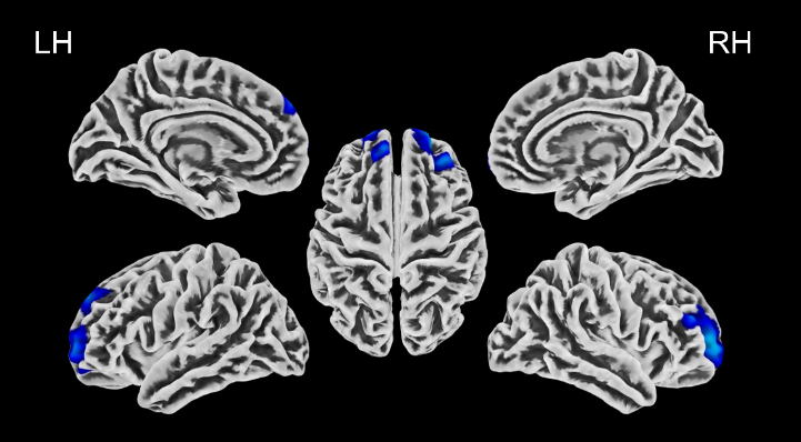
图1与健康对照组相比,视神经脊髓炎谱系疾病患者双侧额中回喙部和左侧额上回皮质厚度显著降低(P < 0.001,团块水平FDR校正)(图源:Huang et al., Neural Regen Res, 2023)

图2与没有视神经炎临床发作的视神经脊髓炎谱系疾病患者相比,既往有视神经炎临床发作的视神经脊髓炎谱系疾病患者双侧楔叶、上顶叶皮质和距状旁回皮质的皮质厚度较低(P < 0.01,团块水平FDR校正)。(图源:Huang et al., Neural Regen Res, 2023)
左侧额中回喙部皮质厚度与数字符号转换测试评分呈正相关(r = 0.469, P = 0.004, PFDR= 0.060),但与连线测试A评分(r = -0.380, P = 0.022, PFDR = 0.110)和扩展残疾状态量表评分(r = -0.318, P = 0.037, PFDR = 0.093)呈负相关。右侧额中回喙部皮质厚度与数字符号转换测试评分呈正相关(r = 0.457, P = 0.005, PFDR = 0.038),与连线测试A评分(r = -0.356, P = 0.033, PFDR = 0.099)、连线测试B评分(r = -0.340, P = 0.042, PFDR = 0.09)以及扩展残疾状态量表评分(r = -0.336, P = 0.028, PFDR = 0.105)呈负相关。而左侧左额上回皮质厚度则与临床参数无相关性,且显著脑区的皮质厚度与病程无相关性(P > 0.05)。
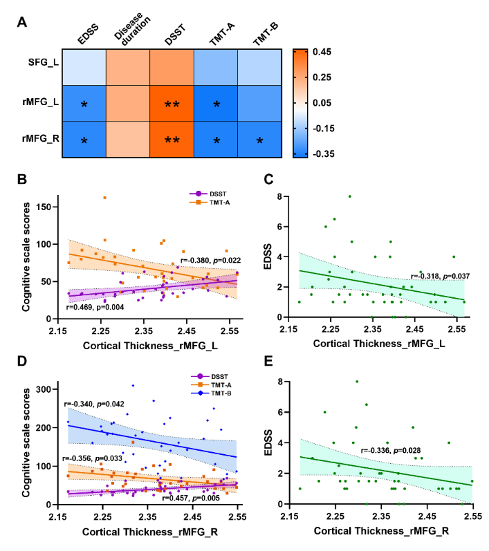
图3皮质厚度平均值与临床参数的相关性(图源:Huang et al., Neural Regen Res, 2023)
在这项研究中,王志远等应用基于表面形态学分析方法量化无明显脑病灶的视神经脊髓炎谱系疾病患者的全脑皮质厚度改变,发现视神经脊髓炎谱系疾病患者双侧额中回喙部的隐匿性皮质萎缩与临床残疾和认知功能(包括工作记忆和执行功能等)相关。进一步亚组分析提示,与没有视神经炎临床发作的视神经脊髓炎谱系疾病患者相比,有视神经炎临床发作的视神经脊髓炎谱系疾病患者的双侧楔叶、上顶叶皮质和距状旁回皮质变薄。已知楔叶参与了基本和高级视觉处理[6],这可能可以解释这些既往有视神经炎临床发作患者发生的长期视觉障碍。此外,楔叶还支持包括记忆在内的非视觉功能[6],还需要未来在脑网络分析方面及其临床相关性的进一步研究。
这项横断面研究的性质使得无法评估视神经脊髓炎谱系疾病不同阶段的多种变化和特征。其次,视神经脊髓炎谱系疾病患者的样本量有限,使得无法根据不同的状态(如认知功能、疾病持续时间以及是否存在脑部病变)将患者进行更细的划分。因此,未来仍需要更大样本量和长期随访的多模态研究以探索视神经脊髓炎谱系疾病的神经机制。
综上,该研究揭示了在无明显脑病灶的视神经脊髓炎谱系疾病患者中,双侧额叶多个脑区皮质厚度的显著降低,并与临床残疾与认知功能具有相关性。这一结果将有助于阐明视神经脊髓炎谱系疾病脑皮质微结构的变化,完善该疾病的成像特点,并有望为临床个体化治疗提供理论基础。
原文链接:https://doi.org/10.4103/1673-5374.371371
参考文献
[1] Hor JY, Asgari N, Nakashima I, et al. Epidemiology of neuromyelitis optica spectrum disorder and its prevalence and incidence worldwide. Front Neurol. 2020;11:501.
[2] Dutra BG, Da Rocha AJ, Nunes RH, et al. Neuromyelitis optica spectrum disorders: spectrum of mr imaging findings and their differential diagnosis. Radiographics. 2018;38(1):169-193.
[3] Charil A, Dagher A, Lerch JP, et al. Focal cortical atrophy in multiple sclerosis: relation to lesion load and disability. Neuroimage. 2007;34(2):509-517.
[4] Treaba CA, Herranz E, Barletta VT, et al. The relevance of multiple sclerosis cortical lesions on cortical thinning and their clinical impact as assessed by 7.0-T MRI. J Neurol. 2021;268(7):2473-2481.
[5] Hyun JW, Park G, Kwak K, et al. Deep gray matter atrophy in neuromyelitis optica spectrum disorder and multiple sclerosis. Eur J Neurol. 2017;24(2):437-445.
[6] Palejwala AH, Dadario NB, Young IM, et al. Anatomy and white matter connections of the lingual gyrus and cuneus. World Neurosurg. 2021;151:e426-e437.
第一作者:黄楚欣,中南大学湘雅二医院放射科医师
通讯作者:王志远,湖南省肿瘤医院/中南大学湘雅医学院附属肿瘤医院超声科主任医师

刘军,教授,中南大学湘雅二医院放射科主任医师
卢伟,中南大学湘雅二医院神经内科主任医师