中国神经再生研究(英文版) ›› 2024, Vol. 19 ›› Issue (4): 811-817.doi: 10.4103/1673-5374.382225

• 综述:脊髓损伤修复保护与再生 • 上一篇    下一篇

利用体内成像解读神经元对损伤的反应:基于脊髓损伤治疗的紧迫性


  • 出版日期:2024-04-15 发布日期:2023-09-15

In vivo imaging of the neuronal response to spinal cord injury: a narrative review

Junhao Deng1, 2, 3, #, Chang Sun1, 2, 4, #, Ying Zheng5, Jianpeng Gao1, 2, Xiang Cui1, 2, Yu Wang6, 7, Licheng Zhang1, 2, *, Peifu Tang1, 2   

  1. 1Department of Orthopedics, The Fourth Medical Center of Chinese PLA General Hospital, Beijing, China; 2National Clinical Research Center for Orthopedics, Sports Medicine and Rehabilitation, Beijing, China; 3School of Life Sciences, Tsinghua University, Beijing, China; 4Department of Orthopedics, Air Force Medical Center, PLA, Beijing, China; 5Medical School of Chinese PLA, Beijing, China; 6Institute of Orthopedics, The First Medical Center, Chinese PLA General Hospital, Beijing, China; 7Key Lab of Regenerative Medicine in Orthopedics, Key Laboratory of Musculoskeletal Trauma and War Injuries PLA, Beijing, China
  • Online:2024-04-15 Published:2023-09-15
  • Contact: Licheng Zhang, PhD, zhanglcheng301@163.com.
  • Supported by:
    This work was supported by the National Natural Science Foundation of China, No. 82272478 (to PT).

摘要:

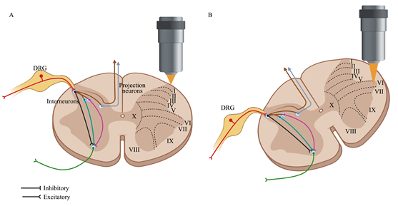
解读脊髓损伤后的神经元反应对于探索脊髓损伤治疗的潜在策略具有重要的意义。然而,目前仍缺乏探索脊髓损伤后神经元对损伤的反应,其部分原因是缺乏合适的工具。传统的电生理学在观察兴奋细胞生物电活动时存在攻击性和侵入性,且无法识别细胞类型或精确的空间位置,也无法进行长期观察。目前,新出现的体内成像和适当标记方法为观察中枢神经系统正常或损伤状态下神经元的动态过程提供了新的途径。此次综述首先对小鼠脊髓进行体内成像,重点是观察脊髓神经元的最新手段进行回顾,分析了脊髓感觉及运动神经元对脊髓损伤的动态生物学反应,总结对比了作为探索脊髓损伤神经元反应的研究手段,并阐明体内成像相较传统神经科学检查的优势,最后还明确了脊髓神经元成像的接下来所面临的挑战和可能的解决方案。

https://orcid.org/0000-0002-1024-4098 (Licheng Zhang)

关键词: 体内成像, 双光子显微镜, 神经元反应, 中枢神经系统, 脊髓, 脊髓损伤, 背角神经元, 前角神经元, 背根神经节, 钙成像

Abstract: Deciphering the neuronal response to injury in the spinal cord is essential for exploring treatment strategies for spinal cord injury (SCI). However, this subject has been neglected in part because appropriate tools are lacking. Emerging in vivo imaging and labeling methods offer great potential for observing dynamic neural processes in the central nervous system in conditions of health and disease. This review first discusses in vivo imaging of the mouse spinal cord with a focus on the latest imaging techniques, and then analyzes the dynamic biological response of spinal cord sensory and motor neurons to SCI. We then summarize and compare the techniques behind these studies and clarify the advantages of in vivo imaging compared with traditional neuroscience examinations. Finally, we identify the challenges and possible solutions for spinal cord neuron imaging.

Key words: anterior horn neurons, calcium imaging, central nervous system, dorsal horn neurons,  , dorsal root ganglion, in vivo imaging, neuronal response, spinal cord injury, spinal cord, two-photon microscopy