中国神经再生研究(英文版) ›› 2026, Vol. 21 ›› Issue (1): 365-376.doi: 10.4103/NRR.NRR-D-24-00381

• 原著:脊髓损伤修复保护与再生 • 上一篇    下一篇

过表达脑源性神经营养因子的小胶质细胞可促进小鼠脊髓损伤后血管再生和功能恢复


  • 出版日期:2026-01-15 发布日期:2025-04-24

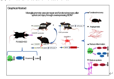
Microglia overexpressing brain-derived neurotrophic factor promote vascular repair and functional recovery in mice after spinal cord injury

Fanzhuo Zeng1, 2, 3, Yuxin Li4 , Xiaoyu Li4 , Xinyang Gu4 , Yue Cao4 , Shuai Cheng1 , He Tian5, *, Rongcheng Mei2, *, Xifan Mei1, *   

  1. 1 Department of Orthopedics, The Third Affiliated Hospital of Jinzhou Medical University, Jinzhou, Liaoning Province, China;  2 Department of Orthopedics, Xiangyang Central Hospital, Affiliated Hospital of Hubei University of Arts and Science, Xiangyang, Hubei Province, China;  3 Department of Neurobiology, School of Basic Medical Sciences, Guangdong-Hong KongMacao Greater Bay Area Center for Brain Science and Brain-Inspired Intelligence, Southern Medical University, Guangzhou, Guangdong Province, China;  4 Institute for Translational Brain Research, State Key Laboratory of Medical Neurobiology, MOE Frontiers Center for Brain Science, Fudan University, Shanghai, China;  5 Liaoning Provincial Collaborative Innovation Center for Medical Testing and Drug Research, Jinzhou Medical University, Jinzhou, Liaoning Province, China
  • Online:2026-01-15 Published:2025-04-24
  • Contact: He Tian, PhD, tianhe@jzmu.edu.cn; Rongcheng Mei, PhD, meirch@163.com; Xifan Mei, PhD, meixifan@jzmu.edu.cn.
  • Supported by:
    This work was supported by the National Natural Science Foundation of China, Nos. 82072165 and 82272256 (both to XM); and the Key Project of Xiangyang Central Hospital, No. 2023YZ03 (to RM).

摘要:

脊髓损伤是一种严重的中枢神经系统创伤性疾病,目前的治疗选择仍然非常有限。小胶质细胞是中枢神经系统中的关键免疫细胞,在脊髓损伤恢复过程中发挥着重要作用。为了研究小胶质细胞在脊髓损伤中的作用,实验在小鼠脊髓损伤前14天开始使用胶质细胞刺激因子1受体抑制剂PLX5622清除小胶质细胞,并在小鼠脊髓损伤后28天内继续清除小胶质细胞。结果发现持续清除小胶质细胞会导致脊髓损伤后病变面积的增大,下调脑源性神经营养因子的表达,并且会导致小鼠脊髓损伤后肢运动恢复能力的下降。因此,实验构建了小胶质细胞条件性过表达脑源性神经营养因子的小鼠,即CX3CR1 creER-/+:LSL-BDNF-/+-tdTomato(CX3:BDNF)。研究发现在小胶质细胞中过表达脑源性神经营养因子促进了脊髓损伤后小鼠后肢运动功能的恢复和血管生成。在脊髓损伤的急性期,在小胶质细胞中过表达 BDNF 可减少炎症因子的产生和神经元的凋亡。通过使用特异性标记小胶质细胞的转基因小鼠TMEM119 creER-/+:LSL-BDNF-+-tdTomato和在CX3:BDNF小鼠中使用一种不通过血脑屏障的胶质细胞刺激因子1受体抑制剂PLX73086,提示主要是小胶质细胞过表达脑源性神经营养因子产生了保护作用,而不是巨噬细胞过表达的脑源性神经营养因子发挥了保护作用。总之,实验证明靶向小胶质细胞过表达脑源性神经营养因子是促进脊髓损伤患者运动功能恢复的一种非常有前景的治疗策略。

https://orcid.org/0000-0002-4452-8005 (Xifan Mei)

关键词: 血管生成, 凋亡, 脑源性神经营养因子, 集落刺激因子1受体, 炎症, 小胶质细胞, 运动功能, 脊髓损伤, 血管内皮生长因子

Abstract: Spinal cord injury represents a severe form of central nervous system trauma for which effective treatments remain limited. Microglia is the resident immune cells of the central nervous system, play a critical role in spinal cord injury. Previous studies have shown that microglia can promote neuronal survival by phagocytosing dead cells and debris and by releasing neuroprotective and anti-inflammatory factors. However, excessive activation of microglia can lead to persistent inflammation and contribute to the formation of glial scars, which hinder axonal regeneration. Despite this, the precise role and mechanisms of microglia during the acute phase of spinal cord injury remain controversial and poorly understood. To elucidate the role of microglia in spinal cord injury, we employed the colony-stimulating factor 1 receptor inhibitor PLX5622 to deplete microglia. We observed that sustained depletion of microglia resulted in an expansion of the lesion area, downregulation of brain-derived neurotrophic factor, and impaired functional recovery after spinal cord injury. Next, we generated a transgenic mouse line with conditional overexpression of brain-derived neurotrophic factor specifically in microglia. We found that brain-derived neurotrophic factor overexpression in microglia increased angiogenesis and blood flow following spinal cord injury and facilitated the recovery of hindlimb motor function. Additionally, brain-derived neurotrophic factor overexpression in microglia reduced inflammation and neuronal apoptosis during the acute phase of spinal cord injury. Furthermore, through using specific transgenic mouse lines, TMEM119, and the colony-stimulating factor 1 receptor inhibitor PLX73086, we demonstrated that the neuroprotective effects were predominantly due to brain-derived neurotrophic factor overexpression in microglia rather than macrophages. In conclusion, our findings suggest the critical role of microglia in the formation of protective glial scars. Depleting microglia is detrimental to recovery of spinal cord injury, whereas targeting brain-derived neurotrophic factor overexpression in microglia represents a promising and novel therapeutic strategy to enhance motor function recovery in patients with spinal cord injury.

Key words: angiogenesis, apoptosis, brain-derived neurotrophic factor, colony stimulating factor 1 receptor, inflammation, microglia, motor function, spinal cord injury, vascular endothelial growth factor