中国神经再生研究(英文版) ›› 2025, Vol. 20 ›› Issue (5): 1455-1466.doi: 10.4103/NRR.NRR-D-23-00794

• 原著:脑损伤修复保护与再生 • 上一篇    下一篇

脑缺血再灌注损伤中内质网的应激和自噬:PERK可能成为靶点?


  • 出版日期:2025-05-15 发布日期:2024-10-31

Endoplasmic reticulum stress and autophagy in cerebral ischemia/reperfusion injury: PERK as a potential target for intervention

Ju Zheng1, 2, # , Yixin Li 3, # , Ting Zhang1 , Yanlin Fu1 , Peiyan Long1 , Xiao Gao1 , Zhengwei Wang1 , Zhizhong Guan1 , Xiaolan Qi 1, 4 , Wei Hong1, 4, * , Yan Xiao1, 4, *   

  1. 1 Key Laboratory of Endemic and Ethnic Diseases, Ministry of Educatton & Key Laboratory of Medical Molecular Biology of Guizhou Province, Guizhou Medical University, Guiyang, Guizhou Province, China;  2 Guizhou Center for Disease Control and Preventton, Guiyang, Guizhou Province, China;  3 Department of Histology and Embryology, Guizhou Medical University, Guiyang, Guizhou Province, China;  4 Collaborattve Innovatton Center for Preventton and Control of Endemic and Ethnic Regional Diseases Co-constructed by the Province and Ministry, Guiyang, Guizhou Province, China
  • Online:2025-05-15 Published:2024-10-31
  • Contact: Yan Xiao, PhD, xiaoyanhonan@hotmail.com; Wei Hong, PhD, hongwei@gmc.edu.cn.
  • Supported by:
    This work was supported by the Nattonal Natural Science Foundatton of China, Nos. 82260245 (to YX), 81660207 (to YX), 81960253 (to YL), 82160268 (to YL), U1812403 (to ZG); Science and Technology Projects of Guizhou Province, Nos. [2019]1440 (to YX), [2020]1Z067 (to WH); Culttvatton Foundatton of Guizhou Medical University, No. [20NSP069] (to YX); Excellent Young Talents Plan of Guizhou Medical University, No. (2022)101 (to WH).

摘要:

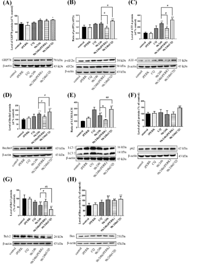
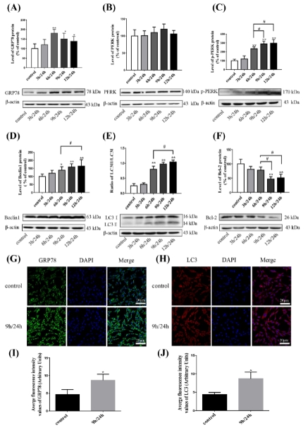
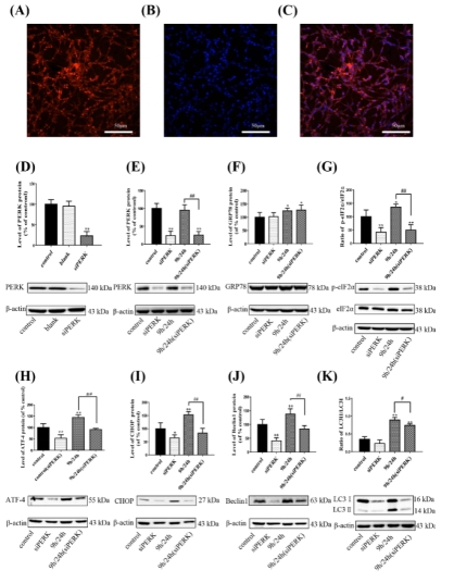
研究表明,未折叠蛋白反应和内质网应激激活在严重脑缺血再灌注损伤中起着至关重要的作用。脑缺血后数小时内即可发生细胞自噬,但内质网应激和自噬之间的关系尚不清楚。为此,实验对PC12细胞和原代神经元行氧糖剥夺复氧损伤以模拟脑缺血再灌注损伤。结果表明,随着氧糖剥夺时间延长,内质网应激蛋白激酶R样内质网激酶(PERK/真核细胞翻译起始因子eIF2α-激活转录因子4ATF4-C/EBP 同源蛋白(CHOP)通路随之激活,并促进神经元的凋亡以及自噬的过度激活。而抑制内质网应激或敲低PERK基因可显著减弱由氧糖剥夺复氧诱导的过度自噬和神经元凋亡,这表明自噬和内质网应激存在关联,提示PERK是调节过度自噬的重要靶点。此外,以特异性自噬抑制剂氯喹阻断自噬可加剧内质网应激诱导的细胞凋亡,表明适当的自噬可在脑缺血再灌注后的神经元损伤中起保护作用。综上说明,脑缺血再灌注损伤可引发神经元内质网应激并促进其自噬,且PERK是抑制脑缺血再灌注损伤自噬过度激活的可能靶点

https://orcid.org/0009-0004-6220-9608 (Yan Xiao); https://orcid.org/0000-0002-3317-9401 (Wei Hong) 

关键词:

内质网应激, PERK, eIF2α, ATF4, C/EBP 同源蛋白, 自噬, 凋亡, 脑缺血再灌注

Abstract:

Several studies have shown that activation of unfolded protein response and endoplasmic reticulum (ER) stress plays a crucial role in severe cerebral ischemia/reperfusion injury. Autophagy occurs within hours after cerebral ischemia, but the relationship between ER stress and autophagy remains unclear. In this study, we established experimental models using oxygen-glucose deprivation/reoxygenation in PC12 cells and primary neurons to simulate cerebral ischemia/reperfusion injury. We found that prolongation of oxygen-glucose deprivation activated the ER stress pathway protein kinase-like endoplasmic reticulum kinase (PERK)/eukaryotic translation initiation factor 2 subunit alpha (eIF2α)-activating transcription factor 4 (ATF4)-C/EBP homologous protein (CHOP), increased neuronal apoptosis, and induced autophagy. Furthermore, inhibition of ER stress using inhibitors or by siRNA knockdown of the PERK gene significantly attenuated excessive autophagy and neuronal apoptosis, indicating an interaction between autophagy and ER stress and suggesting PERK as an essential target for regulating autophagy. Blocking autophagy with chloroquine exacerbated ER stress-induced apoptosis, indicating that normal levels of autophagy play a protective role in neuronal injury following cerebral ischemia/reperfusion injury. Findings from this study indicate that cerebral ischemia/ reperfusion injury can trigger neuronal ER stress and promote autophagy, and suggest that PERK is a possible target for inhibiting excessive autophagy in cerebral ischemia/reperfusion injury. 

Key words:

apoptosis, ATF4, autophagy, C/EBP homologous protein, cerebral ischemia/reperfusion injury, eIF2α, endoplasmic reticulum stress,  PERK